
|
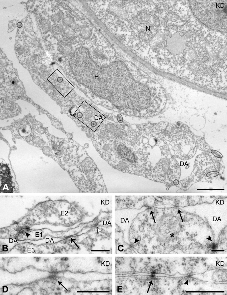
Fig. 6 Altered positions of endothelial cell-endothelial cell (EC-EC) cell junctions in the axial vascular area of the trunk of Egfl7 knockdown (KD) embryos at 24 hours postfertilization (hpf). A: Higher magnification of dorsal aorta (DA) boxed in Figure 5. ECs are linked by tight junctions (encircled) and form a flattened, ring-like endothelium. Ectopic cell-cell junctions (boxes) are positioned within the luminal EC plasma membranes, crossing the obliterated vessel lumen. B,C: Higher magnifications of the ectopic cell junctions in the boxed areas of A. B: From a section consecutive to the section shown in A. A dorsally located EC (E1) is connected to another dorsal EC (E2) by a regular tight junction (arrowhead), and across the collapsed vessel lumen (DA) to a ventrally located EC (E3) by an aberrantly positioned gap junction (arrow) and a tight junction (open arrowhead). C: An EC (*) is linked by regular tight junctions to its lateral neighbors (arrowheads) and by two aberrantly positioned tight junctions (arrows) to an EC at the opposite side of the vessel. D: Example of an ectopic adherens junction (arrow) linking the luminal plasma membrane domains of opposed ECs in the DA. E: Tight junction (arrowhead) and desmosome (arrow) linking the basal plasma membrane domains of ECs. H, hypochord; N, notochord. Scale bars = 2 μm in A, 200 nm in B-D; 500 nm in E.