
|
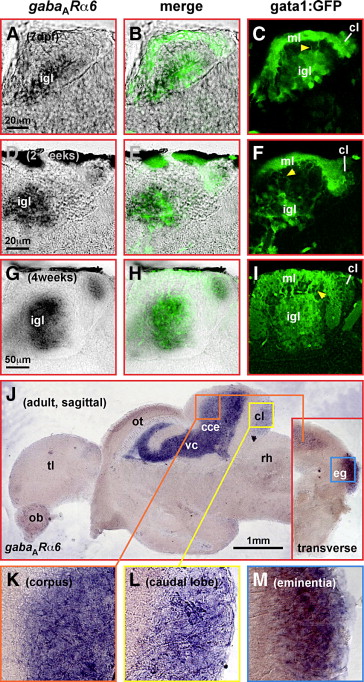
Fig. S1 Sagittal brain sections of 1-, 2- and 4-week-old gata1:GFP transgenic larvae show that gabaARα6-expression (A, D, G) is localized to the internal granule cells layer (igl) of the corpus cerebelli and the granule cell containing caudal lobe (cl), respectively. The same is true for GFP-expression (C, F, I) at these different juvenile stages co-localizing to gabaARα6-expression (B, E, H). Moreover, GFP-containing parallel fibers of gata1:GFP cells extending from the internal granule cell layer (igl) into the molecular layer (ml) can be identified (C, F, I yellow arrowhead). In the adult brain, sagittal sections reveal gabaARα6-expression in the granule cell layer of the corpus cerebelli (J, K) and the caudal lobe (J, L), whereas transverse sections identify additional expression in the granule cell layer of the eminentia granularis (J inset, M). Abbr.: cce, corpus cerebelli; cl, caudal lobe; eg, eminentia granularis; igl, internal granule cell layer; MHB, midbrain–hindbrain boundary; ml, molecular layer; ob, olfactory bulb; ot, optic tectum; rh, rhombencephalon; tl, telencephalon; vc, valvula cerebelli.
Reprinted from Developmental Biology, 313(1), Volkmann, K., Rieger, S., Babaryka, A., and Köster, R.W., The zebrafish cerebellar rhombic lip is spatially patterned in producing granule cell populations of different functional compartments, 167-180, Copyright (2008) with permission from Elsevier. Full text @ Dev. Biol.