
|
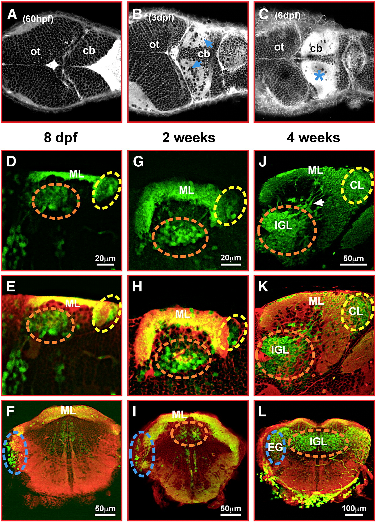
Fig. 5 gata1:GFP granule cells contribute to the corpus cerebelli, the eminentia granularis and the cerebellar caudal lobe. (A–C) Dorsal views of the embryonic and larval zebrafish cerebellum stained with Bodipy FL C5-ceramide. Staining of cellular membranes, intense in neuropil (blue arrows and asterisk), was recorded by optical sectioning using laser scanning microscopy. (D, E, G, H, J, K) sagittal sections and (F, I, L) transverse sections through the cerebellum of 8 dpf (D–F), 2 weeks (G–I) and 4 weeks (J–L) old gata1:GFP larval and juvenile fish. GFP-expression (D, G, J) was visualized by immunohistochemistry followed by red fluorescent counterstaining with Bodipy 630/650-X (E, F, H, I, K, L) and image recording using laser scanning confocal microscopy. A dashed circle marks the forming IGL of the corpus cerebelli (orange), the eminentia granularis (blue) and the caudal lobe (yellow), respectively. Note GFP-expressing granule cells still appearing to migrate into the IGL of the corpus cerebelli at 4 weeks (J, white arrow). Abbr.: cb, cerebellum; CL, caudal lobe; EG, eminentia granularis; IGL, internal granule cell layer; ML, molecular layer; ot, optic tectum.
Reprinted from Developmental Biology, 313(1), Volkmann, K., Rieger, S., Babaryka, A., and Köster, R.W., The zebrafish cerebellar rhombic lip is spatially patterned in producing granule cell populations of different functional compartments, 167-180, Copyright (2008) with permission from Elsevier. Full text @ Dev. Biol.