
|
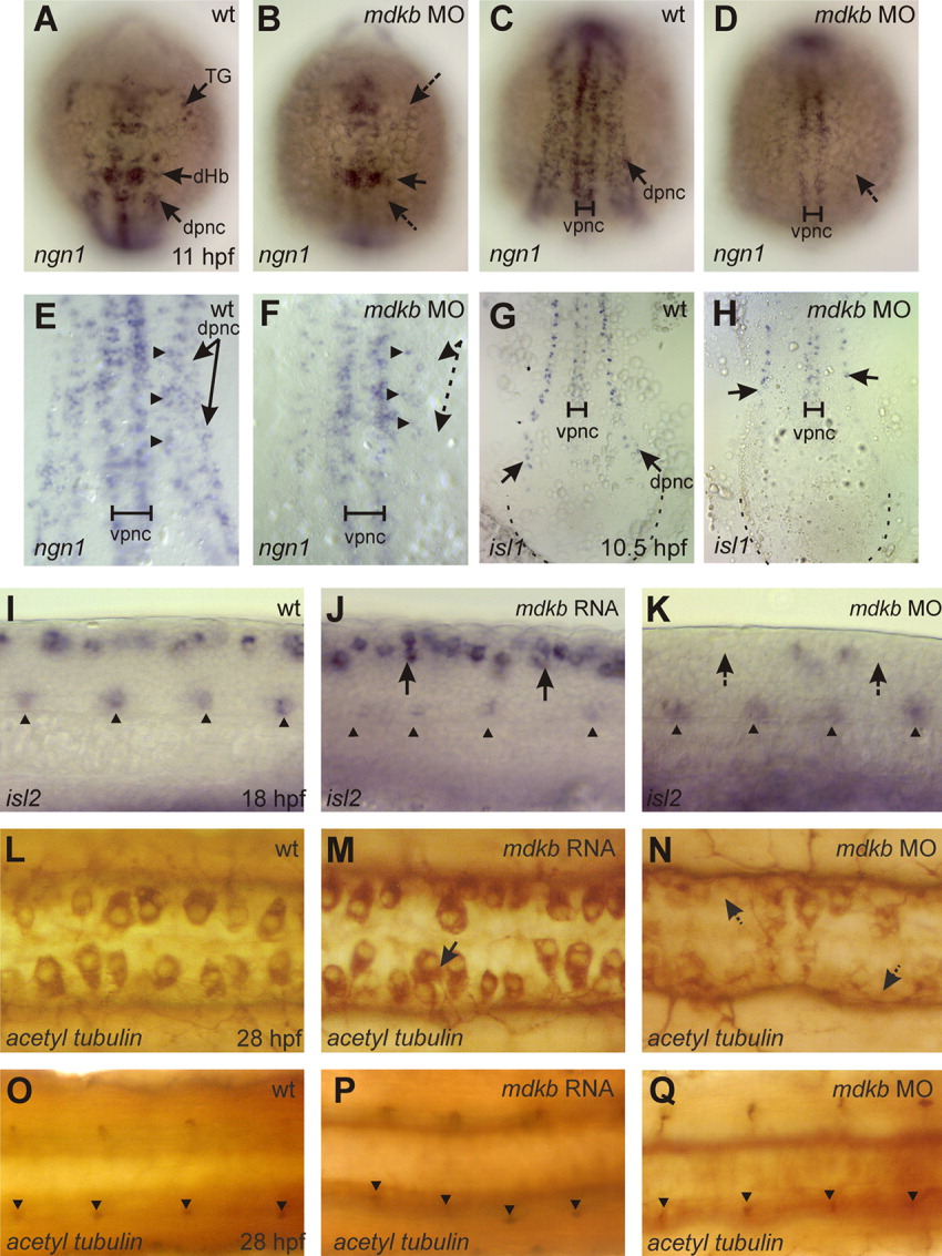
Fig. 6 mdkb regulates formation of sensory neurons. A-H: Dorsal views of trunk regions in noninjected (A,C,E,G) and mdkb Morpholino (MO) -injected embryos (B,D,F,H) at the one- to three-somite stage showing ngn1 (A-F) and isl1 (G,H) expression. Note that the embryo in H is slightly delayed due to MO injection. Dorsal proneural cells (dpnc) are reduced in injected embryos at mid-hindbrain (arrows in A,B) and trunk level (arrows in C-H), while number of ventral proneural cells (vpnc) is not affected. Note remaining interneurons (arrowheads), but absent sensory neurons (arrows) in mdkb morphants (in E,F). I-K: Lateral views of trunk regions in embryos at the 18-somite stage showing isl2 expression in ventrally located motor neurons (arrowheads) and dorsal sensory Rohon-Beard (RB) neurons (arrows). Embryos injected with 150 pg of mdkb RNA (J) show enhanced numbers of dorsal RB, while motor neurons are not affected. In contrast, RB are reduced in mdkb MO-injected embryos (K). L-Q: Dorsal views of embryos at 28 hours postfertilization (hpf) immunostained with an antibody against acetylated tubulin. L-N: Focal planes at the level of dorsal sensory neurons. O-Q: Focal planes of the same embryos at the level of ventral motor neurons. Note increased RB cell number in mdkb RNA injected embryos (arrow in M) and reduced RB cell number in mdkb MO-injected embryos (arrow in N), while number of motor neurons is not affected (arrowheads in M-O). DHb, dorsal hindbrain; TG, trigeminal ganglia; VMb, ventral midbrain.