
|
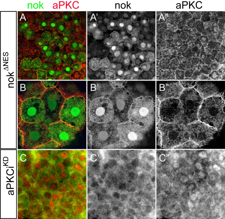
Fig. 6 nok and aPKCi are not sufficient to tether each other within the nucleus. Images are reconstructions of confocal Z-stack sections imaged on late gastrula stage whole mounts. Myc and aPKC stainings are false-colored. A,B: Embryos expressing high levels of HisMyc-tagged nokΔNES were counterstained with anti aPKC. A,A′: Within DL cells, high levels of nuclear HisMyc-tagged nokΔNES are present whereas aPKC localizes to outer cell membranes and no increased levels of nuclear aPKC are detectable (A“). B,B′: Within epithelial EVL cells, HisMyc-tagged nokΔNES is at outer cell membranes and within the nucleus. B”: aPKC is predominantly associated with outer cell membranes. C: Embryos overexpressing HisMyc-tagged nokwt (green) and non-tagged aPKCiKD (red), whereas nokwt is absent from the nucleus (C′), aPKCiKD is nuclear. All images are apical views onto the tissue.