
Image
|
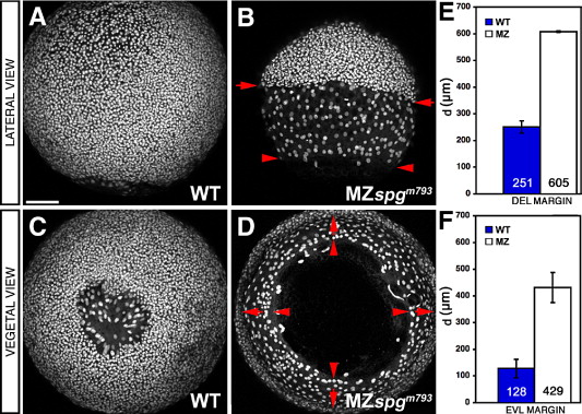
Figure Caption
Fig. 2 Epiboly delay of the DEL and EVL. (A–D) Confocal z-projections of Sytox green stained embryos at 85% epiboly stage. MZspgm739 embryos show a strong DEL (arrows) and EVL (arrowheads) retardation. (A, B) Lateral and (C, D) vegetal views. (E) Measurements of the mean diameter “d” of diametral DEL margins: WT = 251 ± 26 μm, MZspgm793 = 605 ± 6 µm, p = 2 · 10- 08. (F) Measurements of the mean diameter “d” of diametral EVL margins: WT = 128 ± 31 μm, MZspgm739 = 429 ± 13 µm (p = 1 · 10- 06). Scale bar: 100 μm.
Figure Data
Acknowledgments
This image is the copyrighted work of the attributed author or publisher, and
ZFIN has permission only to display this image to its users.
Additional permissions should be obtained from the applicable author or publisher of the image.
Reprinted from Developmental Biology, 315(1), Lachnit, M., Kur, E., and Driever, W., Alterations of the cytoskeleton in all three embryonic lineages contribute to the epiboly defect of Pou5f1/Oct4 deficient MZspg zebrafish embryos, 1-17, Copyright (2008) with permission from Elsevier. Full text @ Dev. Biol.