
Image
|
Figure Caption
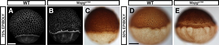
Fig. S6 Maternal contribution to the YCL phenotype. (A, B) Alexa488-Phalloidin staining. (C–E) Anti-β-Tubulin antibody immunohistochemistry with secondary antibody/HRP/DAB detection. (A–C) 75% epiboly, (D, E) 50% epiboly embryos. (A, D) WT; (B, C, E) Mspgm793. (E) Microtubule track formation in Mspgm793 embryos is slightly distorted, but microtubule free patches are not observed. Scale bars: 100 μm.
Figure Data
Acknowledgments
This image is the copyrighted work of the attributed author or publisher, and
ZFIN has permission only to display this image to its users.
Additional permissions should be obtained from the applicable author or publisher of the image.
Reprinted from Developmental Biology, 315(1), Lachnit, M., Kur, E., and Driever, W., Alterations of the cytoskeleton in all three embryonic lineages contribute to the epiboly defect of Pou5f1/Oct4 deficient MZspg zebrafish embryos, 1-17, Copyright (2008) with permission from Elsevier. Full text @ Dev. Biol.