
|
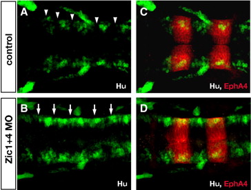
Fig. S3 Zic1 and Zic4-deficient embryos lack normal segmental patterns of neuronal differentiation in the hindbrain. (A,B) Longitudinal confocal sections through the dorsal hindbrain of Hu immunolabeling (green, labels post-mitotic neurons), and (C,D) of double labeling with antibodies to Hu (green) and EphA4 (red, labels r3 and r5). In 24 hpf control embryos (A,C) post-mitotic neurons are clustered in the center of rhombomeres and absent from rhombomere boundaries (white arrowheads in A), while in Zic1 + 4 morphants (B,D) segmental pattern of Hu labeling is disrupted with ectopic differentiation of Hu-positive neurons in boundary regions (white arrows in B).
Reprinted from Developmental Biology, 314(2), Elsen, G.E., Choi, L.Y., Millen, K.J., Grinblat, Y., and Prince, V.E., Zic1 and Zic4 regulate zebrafish roof plate specification and hindbrain ventricle morphogenesis, 376-392, Copyright (2008) with permission from Elsevier. Full text @ Dev. Biol.