
Fig. 6
|
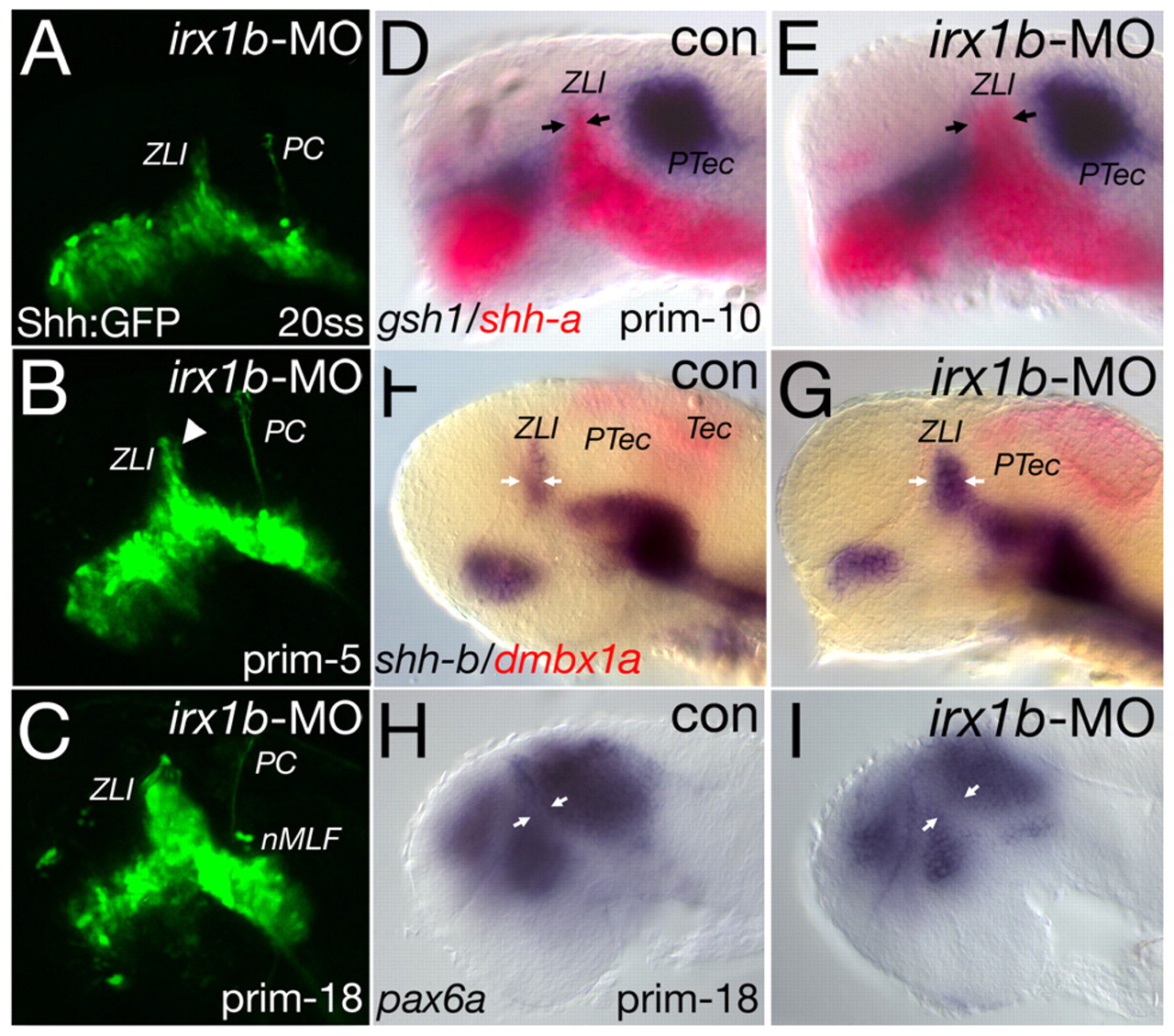
Fig. 6 Irx1b sets the posterior boundary of the ZLI. Confocal microscopy analysis of transgenic shh-GFP; irx1b-morpholino (MO) embryos (A-C) and in situ-hybridisation analysis of irx1b-MO embryos (D-I); markers and stages are indicated. (A) irx1b-MO-injected embryos resemble control (con) embryos at the same stage (compare with Fig. 3). (B) At 26 hpf, cells located posterior to the ZLI start to express Shh-GFP ectopically (arrowhead), whereas the posterior commissure (PC) and the nucleus of the medio-longitudinal fascicle (nMLF) form normally. (C) At 32 hpf, the Shh-GFP expression at the ZLI is broadened compared with control-injected embryos (see Fig. 3). (D,E) At prim-10, the Shh expression domain at the ZLI is broadened at the expense of the thalamus (arrows). (F,G) Similarly, the expression domain of shhb expands posteriorly in irx1b morphants (arrows). (D-G) Interestingly, the expression of markers of the pretectum, such as gsh1 and dmbx1a, is unaltered. (H,I) The broadening of the ZLI is also visible by an in situ hybridisation for pax6a, revealing a much wider gap between the prethalamus and the remaining thalamic-pretectal tissue at prim-18 (32 hpf; arrows). nMLF, nucleus of the medio-longitudinal fascicle; PC, posterior commissure; prim, primordium stage; PTec, pretectum; Tec, tectum; ZLI, zona limitans intrathalamica.