
Image
|
Figure Caption
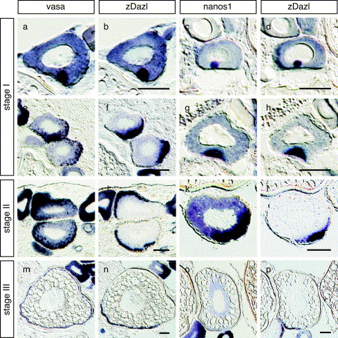
Fig. 1 The distributions of germ plasm RNAs during oogenesis. In situ hybridization of serial sections of the zebrafish ovary. Distributions of vasa and dazl RNAs (a, b, e, f, i, j, m and n) and nanos1 and dazl RNAs (c, d, g, h, k, l, o and p) are compared in the serial sections of oocyte at stages I (a–h), II (i–j), and III (m–p). Sections were cut at a thickness of 10 μm. Scale bars, 50 μm
Figure Data
Acknowledgments
This image is the copyrighted work of the attributed author or publisher, and
ZFIN has permission only to display this image to its users.
Additional permissions should be obtained from the applicable author or publisher of the image.
Reprinted from Mechanisms of Development, 124(4), Kosaka, K., Kawakami, K., Sakamoto, H., and Inoue, K., Spatiotemporal localization of germ plasm RNAs during zebrafish oogenesis, 279-289, Copyright (2007) with permission from Elsevier. Full text @ Mech. Dev.