
Image
|
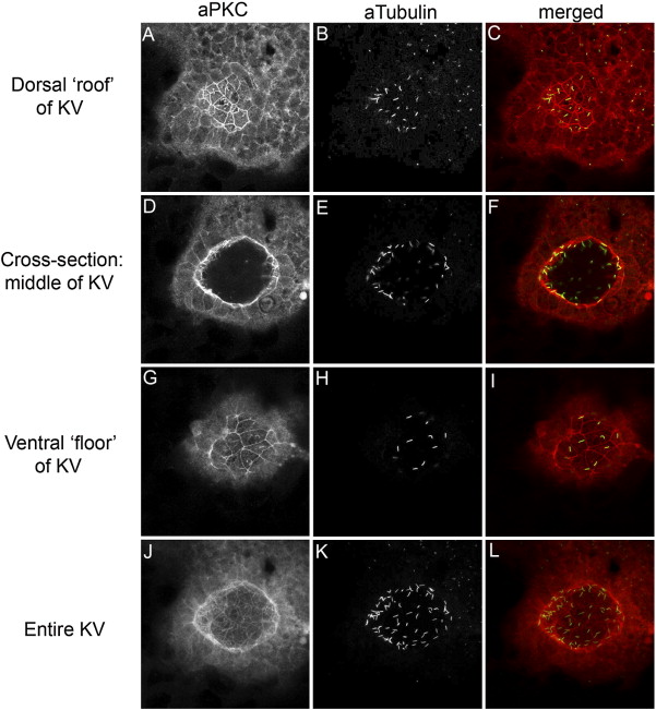
Figure Caption
Fig. S2 aPKC is a marker of KV cells. (A–L) Confocal microscopy of double fluorescent immunostaining in a wild-type embryo using anti-aPKC (red) and anti-acetylated Tubulin (green) antibodies shows that aPKC localizes to the apical membrane of ciliated KV cells. Confocal cross-sections through the dorsal ‘roof’ (A–C), mid-section (D–F) and ventral ‘floor’ (G–I) of KV demonstrate this apical staining on the surfaces lining the KV lumen. Additionally, each of these surfaces is ciliated. (J–L) The sum of all confocal sections through KV.
Acknowledgments
This image is the copyrighted work of the attributed author or publisher, and
ZFIN has permission only to display this image to its users.
Additional permissions should be obtained from the applicable author or publisher of the image.
Reprinted from Developmental Biology, 310(2), Amack, J.D., Wang, X., and Yost, H.J., Two T-box genes play independent and cooperative roles to regulate morphogenesis of ciliated Kupffer's vesicle in zebrafish, 196-210, Copyright (2007) with permission from Elsevier. Full text @ Dev. Biol.