
|
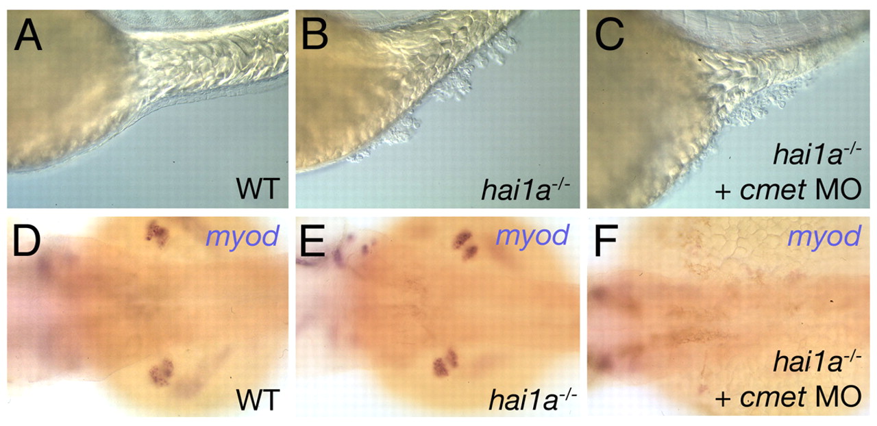
Fig. 8 Inactivation of Met fails to rescue epithelial defects in hai1a mutants. (A-C) Lateral Nomarski images of a wild-type sibling (WT; A), hai1a mutant (B) and met (cmet) MO-injected hai1a mutant (C) at 48 hpf, revealing the presence of epidermal aggregates both in injected (C) and un-injected (B) mutants. (D-F) Dorsal views of the trunk region of embryos shown in A-C, with the pectoral fin muscle labeled for myoD transcripts. The pectoral fin muscle is absent from the met morphant (F), compared with un-injected siblings (D,E), demonstrating that the MO is functional. Notice the presence of normal pectoral fin muscle in the hai1a mutant (E), indicating that Hai1a is dispensable for the Hgf- and Met-dependent migration of fin muscle precursor cells (Haines et al., 2004).