
|
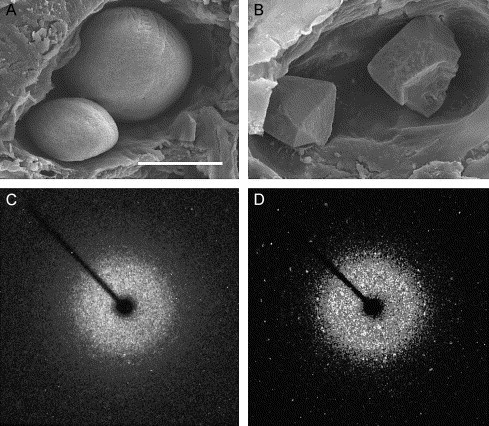
Fig. 7 Calcitic otolith formation in otop1 morphant fish. (A) Scanning electron microscopy of the otic cavity of a 7 dpf uninjected age-matched control showing a smaller utricular (left) and larger saccular otolith. Both otoliths are rounded and cover the entire sensory maculae. (B) Eight-ng MO-1-injected morphant otic cavity at 7 dpf. Otoliths are angular. The sensory epithelium is visible below the utricular otolith. The morphant otoliths resembled inorganic crystals instead of organic calcification. (C) The 360° rotation image of a single crystal X-ray diffraction of a wild-type 7 dpf otolith. Little prominent diffraction pattern is present, indicating that calcium carbonate aragonite crystallites are arranged in a dustlike mosaic pattern across the surface of the otolith. (D) Single crystal X-ray diffraction of a morphant otolith showing that otoliths similar to those above (B) behave as a single crystal. The unit cell derived from this diffraction pattern was consistent with the calcitic polymorph of calcium carbonate. Scale bar indicates 50 μm.
Reprinted from Developmental Biology, 276(2), Hughes, I., Blasiole, B., Huss, D., Warchol, M.E., Rath, N.P., Hurle, B., Ignatova, E., David Dickman, J., Thalmann, R., Levenson, R., and Ornitz, D.M., Otopetrin 1 is required for otolith formation in the zebrafish Danio rerio, 391-402, Copyright (2004) with permission from Elsevier. Full text @ Dev. Biol.