
|
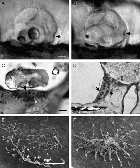
Fig. 5 Morphogenesis of the otocyst and sensory epithelium. (A and B) Lateral view of a 5 dpf wild-type (A) and morphant (B) otocyst. In the wild-type fish, the utricular otolith is smaller and located in the rostral portion of the otocyst, the saccular otolith is larger and located centrally. The semicircular canals and ampullae (arrowhead) are similar in control and in 10 ng MO-1 morphant fish with otolith agenesis. (C) Plastic section through the utricular macula of a 7 dpf wild-type fish (100×) showing development of the hair cells (pale apical cells, arrow) and supporting cells (darker basal cells, arrowhead). (D) Plastic section of a 7 dpf morphant saccular macula showing a similar distribution of cell types. (E and F) Scanning electron micrographs of a wild-type (E) and morphant (F) utricular otolithic membrane at 7 dpf. Removal of the otolith disrupted the wild-type otolithic membrane. Morphant fish formed an otolithic membrane in the absence of the otolith. The otolithic membrane is fibrous and connects the stereocilia of all hair cells in the macula. O, otolith. Scale bars: A–B, 250 μm; C–D, 10 μm; E–F, 10 μm.
Reprinted from Developmental Biology, 276(2), Hughes, I., Blasiole, B., Huss, D., Warchol, M.E., Rath, N.P., Hurle, B., Ignatova, E., David Dickman, J., Thalmann, R., Levenson, R., and Ornitz, D.M., Otopetrin 1 is required for otolith formation in the zebrafish Danio rerio, 391-402, Copyright (2004) with permission from Elsevier. Full text @ Dev. Biol.