
Image
|
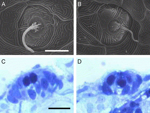
Figure Caption
Fig. 4 Normal formation of the lateral line system in otop1 morphant fish. (A–B) SEM image of a 4 dpf WT (A) and morphant (B) posterior lateral line neuromast. The cupula and kinocilia bundle has formed normally in the morphant animal. (C and D) Four-μm plastic section through a 7 dpf uninjected control (C) and 10 ng MO-1-injected morphant anterior neuromast showing a similar complement of hair cells (pale, apical cells with round nuclei) and supporting cells. Scale bars indicate 10 μm. C and D are stained with Richardson′s stain.
Acknowledgments
This image is the copyrighted work of the attributed author or publisher, and
ZFIN has permission only to display this image to its users.
Additional permissions should be obtained from the applicable author or publisher of the image.
Reprinted from Developmental Biology, 276(2), Hughes, I., Blasiole, B., Huss, D., Warchol, M.E., Rath, N.P., Hurle, B., Ignatova, E., David Dickman, J., Thalmann, R., Levenson, R., and Ornitz, D.M., Otopetrin 1 is required for otolith formation in the zebrafish Danio rerio, 391-402, Copyright (2004) with permission from Elsevier. Full text @ Dev. Biol.