
|
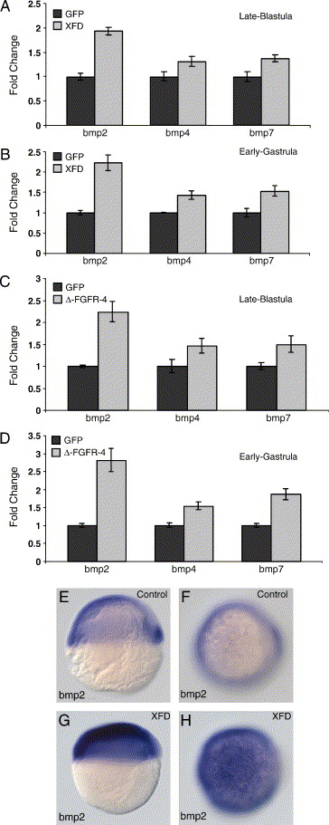
Fig. 5 FGF signaling represses BMP transcription. Real-time PCR analysis of XFD (A and B) and Δ-FGFR-4 (C and D)-microinjected wild-type embryos during late blastula and early gastrula stages. The fold change (y-axis) of the markers bmp2, bmp4, and bmp7 is set relative to control (GFP)-injected embryos. XFD- and Δ-FGFR-4-injected embryos show an increase BMP transcript levels during late blastula stages. (E–H) RNA in situ hybridization of bmp2 expression in control (E and F)- and XFD-injected wild-type embryos (G and H). In XFD-injected embryos, the bmp2 expression domain extends into the dorsal ectoderm. Panels E and G are lateral views of shield stage embryos and F and H are animal pole views of the same embryos.
Reprinted from Developmental Biology, 279(1), Londin, E.R., Niemiec, J., and Sirotkin, H.I., Chordin, FGF signaling, and mesodermal factors cooperate in zebrafish neural induction, 1-19, Copyright (2005) with permission from Elsevier. Full text @ Dev. Biol.