
|
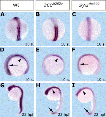
Fig. 7 Expression pattern of grm in WT, fgf8 and shh mutant embryos during somitogenesis and at 22 hpf. Shown are wild-type (A,D,G), aceti282a (B,E,H), and syutbx392 (C, F, I) embryos, stained with a grm probe. Arrows in (D,F) point to grm expression in the anterior mesoderm, and arrow heads in (D,E) to the nascent somites. In (H,I), the arrow heads indicate grm staining in the anterior mesoderm and ventral neural tube, and the arrows point to the restricted grm signal in the posterior region of the mutant embryos. Embryos are oriented with anterior to the top (A–C,G–I), or to the left (D–F). (A–C) are dorsal views, (D–I) lateral views.
Reprinted from Gene expression patterns : GEP, 5(4), Nicoli, S., Gilardelli, C.N., Pozzoli, O., Presta, M., and Cotelli, F., Regulated expression pattern of gremlin during zebrafish development, 539-544, Copyright (2005) with permission from Elsevier. Full text @ Gene Expr. Patterns