
|
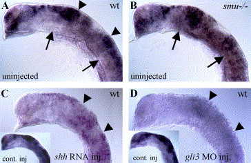
Fig. 3 Hh signaling negatively regulates gli3 expression. (A) In 20-h wild-type embryos, gli3 is expressed regionally in the dorsal neural tube, with strong expression in the tectum and midbrain/hindbrain boundary (arrowheads). (B) gli3 expression is expanded ventrally in smu(smo) mutant embryos that lack Hh pathway activity (arrows). (C) gli3 expression is generally reduced in shh mRNA-injected wild-type embryos (compare to lacZ mRNA-injected embryo in inset). (D) gli3MO injection also led to severely reduced gli3 expression levels (compare to control MO-injected wild-type embryo in inset). All panels show gli3 expression in 20-h zebrafish embryos. Lateral views of head, anterior to the left, eyes and yolk removed.
Reprinted from Developmental Biology, 277(2), Tyurina, O.V., Guner, B., Popova, E., Feng, J., Schier, A.F., Kohtz, J.D., and Karlstrom, R.O., Zebrafish Gli3 functions as both an activator and a repressor in Hedgehog signaling, 537-556, Copyright (2005) with permission from Elsevier. Full text @ Dev. Biol.