
|
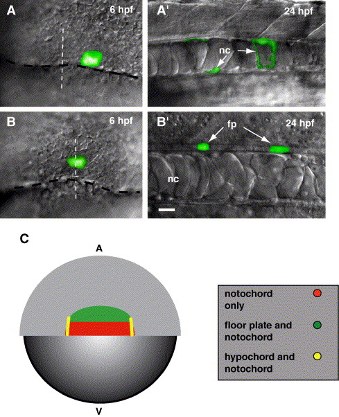
Fig. 1 Midline precursor cells arise from shield domains. Embryos shown in (A′) and (B′) are later stages of the embryos shown in panels A and B, respectively. (A) View of the dorsal margin region of a 6 hpf embryo. Caged fluorescein was photoactivated in 3–5 cells at the margin (black dashed line), near the midline (white dashed line). (A′) Side view of the trunk region of a living embryo at 24 hpf showing labeled notochord (nc) cells. (B) View of the dorsal margin region of a 6 hpf embryo. Cells were labeled approximately 4 cell diameters above the margin (black dashed line) at the midline (white dashed line). (B′) Side view of the trunk region of a living embryo at 24 hpf showing labeled floor plate (fp) cells. (C) Representative schematic of our fate mapping experiments (also see Table 1). The schematic depicts a dorsal view of a 6 hpf embryo, blastoderm is animal (A) and yolk is vegetal (V). We previously showed (Latimer et al., 2002) that hypochord precursor cells (yellow) are located at the lateral edges of the shield. Floor plate precursor cells (green) are located in a region approximately 4–8 cell diameters above the margin. Notochord precursor cells (red) are primarily found directly at the dorsal margin, but were also labeled near hypochord and floor plate precursors. We never labeled floor plate within 4 cell diameters of the blastoderm margin. Scale bar: 20 μm for panels A–B′.
Reprinted from Developmental Biology, 298(2), Latimer, A.J., and Appel, B., Notch signaling regulates midline cell specification and proliferation in zebrafish, 392-402, Copyright (2006) with permission from Elsevier. Full text @ Dev. Biol.