
|
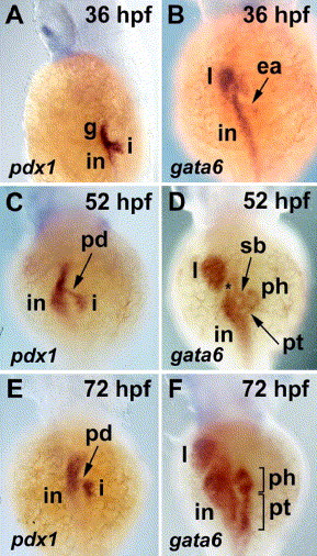
Fig. 2 pdx1 and gata6 expression in the developing zebrafish pancreas and digestive tract; whole mount RNA in situ hybridization, dorsal view. (A, B) At 36 hpf, pdx1 expression in the pancreatic islet, intestine and rostral gut endoderm is evident; gata6 is expressed in the intestine, liver and in the exocrine anlage. (C, D) At 52 hpf, pdx1 is expressed in cells of the endocrine pancreas and a stalk of tissue corresponding to the extrapancreatic duct. Histological sections (not shown) show exocrine cells adjacent to the islet also express pdx1. gata6 is expressed in exocrine cells surrounding the islet that forms the pancreatic head. Tissue of the pancreatic tail is first visible at this stage. (*) pancreatic duct insertion site. (E, F) By 72 hpf, the exocrine pancreas has grown significantly; pdx1 expression in the pancreatic duct is diminished. i: pancreatic islet; in: intestine; g: gut endoderm; l: liver; ea: exocrine anlage; pd: pancreatic duct; sb: swim bladder; ph: pancreatic head; pt: pancreatic tail.
Reprinted from Developmental Biology, 284(1), Yee, N.S., Lorent, K., and Pack, M., Exocrine pancreas development in zebrafish, 84-101, Copyright (2005) with permission from Elsevier. Full text @ Dev. Biol.