
|
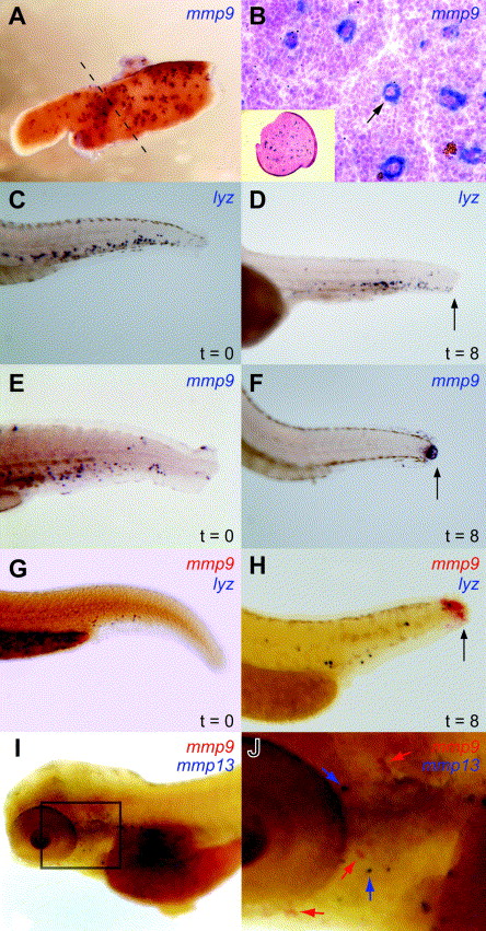
Fig. 3 Further analysis of mmp9-positive cell populations. (A and B) Adult spleen. Whole mount in situ hybridization using a DIG-labeled mmp9 probe of adult zebrafish spleen, either whole (A) or in transverse cross-section (B), showing mmp9 expression (arrow) in the splenic cords of the red pulp of the zebrafish spleen. The inset shows the whole spleen in cross-section. (C–H) Response to trauma. A trauma assay was performed on 48 hpf zebrafish embryos. Embryos either unclipped (t = 0) or 8 h after being tail clipped (t = 8) were subjected to whole mount in situ hybridization with lyz (C and D), mmp9 (E and F), or both lyz and mmp9 (G and H, lyz: blue staining and mmp9: red staining). Embryos are orientated in a lateral view with the anterior to the left and the dorsal side is up. Arrows indicate the migration of cells to the site of trauma. (I and J) Comparison to mmp13+ cell population. Two-color whole mount in situ hybridization performed on wild-type 4 dpf embryos, with both mmp9 (red staining) and mmp13 (blue staining). (J) is an enlargement of the area boxed in (I). Embryos are orientated in a lateral view with the anterior to the left and the dorsal side uppermost, with colored arrows indicating the position of cells expressing each gene.
Reprinted from Gene expression patterns : GEP, 7(1-2), Yoong, S., O'connell, B., Soanes, A., Crowhurst, M.O., Lieschke, G.J., and Ward, A.C., Characterization of the zebrafish matrix metalloproteinase 9 gene and its developmental expression pattern, 39-46, Copyright (2007) with permission from Elsevier. Full text @ Gene Expr. Patterns