
|
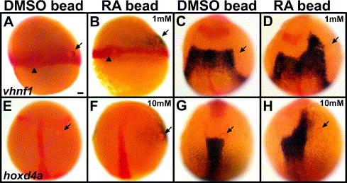
Fig. 5 RA beads can induce posterior hindbrain markers ectopically and prematurely. Embryos with control (A, C, E, G), 1 mM RA (B, D) or 10 mM RA (F, H) beads placed in the margin at 6 hpf (shield stage) and stained in blue for vhnf1 at 7 hpf (65% epiboly) (A, B) or 10.3 hpf (1 s) (C, D) or for hoxd4a at 10 hpf (bud stage) (E, F) or 11.3 hpf (4 s) (G, H). Markers in red are no tail (ntl, Schulte-Merker et al., 1992), which labels the margin and the shield/dorsal midline (A–B) and the notochord (C–F) and also pax2a to label the MHB (C–H) and the early ear primordia (G–H). Dorsal views show anterior to the top. Arrows point to beads. Arrowheads point to the shield/dorsal midline (A–B). (A–D) vhnf1 initiates prematurely on the RA-bead side (B) in 8/8 cases, compared with the control side (0/8) and with control bead embryos (A, 0/8). If bead-implanted embryos are allowed to develop further, to 10.3 hpf, 10/10 control embryos show normal vhnf1 expression (C), and 9/9 embryos with 1 mM RA beads show anterior expansion of vhnf1 on the RA-bead side (D). (E–H) hoxd4a initiates prematurely on the RA-bead side (F) in 7/7 cases, compared with the control side (0/7) and with control bead embryos (E, 0/10). If these embryos are allowed to develop further, to 11.3 hpf, 10/10 control embryos show normal hoxd4a expression (G), and 9/9 embryos with 10 mM RA beads show anterior expansion of hoxd4a on the RA-bead side (H). r3: rhombomere 3 krox-20 expression. Scale bar: 50 μm.
Reprinted from Developmental Biology, 285(2), Maves, L., and Kimmel, C.B., Dynamic and sequential patterning of the zebrafish posterior hindbrain by retinoic acid, 593-605, Copyright (2005) with permission from Elsevier. Full text @ Dev. Biol.