
Image
|
Figure Caption
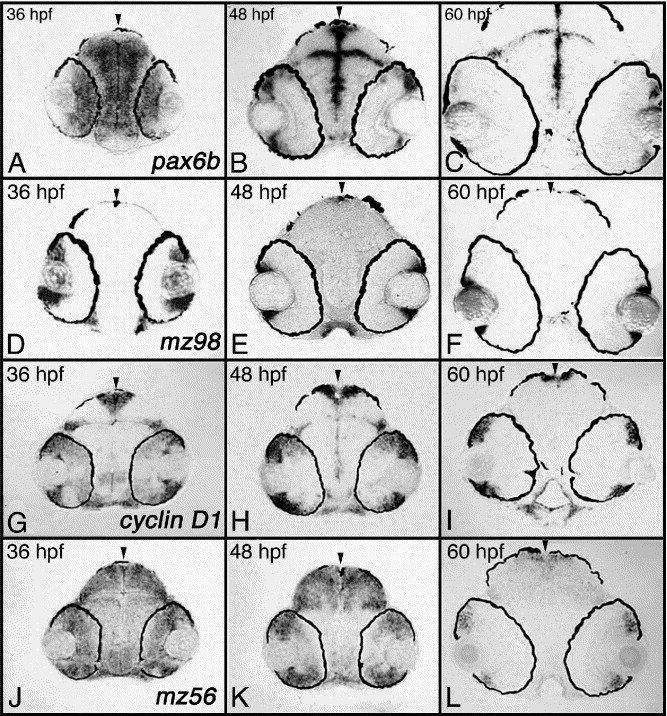
Fig. 5 Marginal zone expression. Shown are examples of transcripts expressed at the marginal zone. (A–C) pax6b, (D–F) mz98, (G–I) cyclin D1, (J–L) mz56. Transcripts were analyzed on plastic sections following whole-embryo in situ hybridization. Arrowheads mark the midline. hpf, hours postfertilization.
Figure Data
Acknowledgments
This image is the copyrighted work of the attributed author or publisher, and
ZFIN has permission only to display this image to its users.
Additional permissions should be obtained from the applicable author or publisher of the image.
Reprinted from Developmental Biology, 293(2), Pujic, Z., Omori, Y., Tsujikawa, M., Thisse, B., Thisse, C., and Malicki, J., Reverse genetic analysis of neurogenesis in the zebrafish retina, 330-347, Copyright (2006) with permission from Elsevier. Full text @ Dev. Biol.