
|
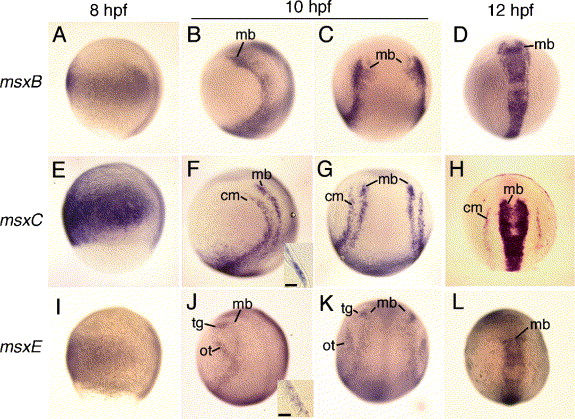
Fig. 1 Expression of msxB, msxC, and msxE. Wild type embryos at 8 hpf (A, E, I), 10 hpf (B, C, F, G, J, K) and 12 hpf (D, H, L) showing msxB expression (A–D), msxC expression (E–H), and msxE expression (I–L). The inset in panel F shows a cross section through the cranial mesoderm (cm) domain of msxC. The inset in panel J shows a cross section through the otic (ot) domain of msxE, where expression is limited to the ectoderm. Scale bars in the insets = 10 μm. Abbreviations: cm, cranial mesoderm; mb, midbrain; ot, otic; tg, trigeminal. Anterior is to the top in all specimens. Panels A, B, E, F, I, J show lateral views with dorsal to the right. Panels C, D, G, H, K, L show dorsal views.
Reprinted from Developmental Biology, 294(2), Phillips, B.T., Kwon, H.J., Melton, C., Houghtaling, P., Fritz, A., and Riley, B.B., Zebrafish msxB, msxC and msxE function together to refine the neural-nonneural border and regulate cranial placodes and neural crest development, 376-390, Copyright (2006) with permission from Elsevier. Full text @ Dev. Biol.