
|
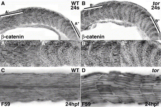
Fig. 4 Somite boundaries form normally, but defects appear later, as tor mutant somitic cells mature into skeletal muscle. Confocal micrographs of β-catenin expression show similar cubiodal morphology of cells at newly formed/forming boundaries in wild type (A, A″) and tor mutant (B, B″) embryos. In wild type embryos, myofibers in anterior somites have extended across the entire myotome (A′), while myofibers in anterior mutant somites have not completed elongation (B′). The bars in Panels A and B indicate the respective regions magnified in the prime and double prime panels. Slow muscle myosin expression (C, D) shows the abnormal morphology of slow muscle in tor mutant embryos; slow fibers in mutant embryos are irregularly spaced, do not extend in a straight trajectory, and boundaries between the myotomes are irregular. Panels are lateral views, anterior to the left. Genotype and stage are indicated on the panels.
Reprinted from Developmental Biology, 287(2), Dill, K.K., and Amacher, S.L., tortuga refines Notch pathway gene expression in the zebrafish presomitic mesoderm at the post-transcriptional level, 225-236, Copyright (2005) with permission from Elsevier. Full text @ Dev. Biol.