
|
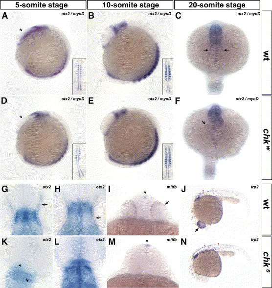
Fig. 6 The specification and differentiation of the RPE do not occur in chk. In situ hybridizations with probes for otx2 (A–H, K, L), mitfb (I, M) and trp2 (J, N) performed in mutant (D–F, K–N) and wt embryos (A–C, G–J) indicate that the RPE is specifically affected in chk. At the 5-somite stage, the expression pattern of otx2 is prematurely downregulated in the eye field of chk compared to wt (arrowheads in panels D and A, respectively). This downregulation resembles the expression pattern of oxt2 in wt embryos at the 10-somite stage (panel B compared to panels D and E). At the 20-somite stage, otx2 is expressed in the RPE of wt embryos (arrows in panel C), weakly expressed in chkw (F) and absent in chks (K). The myogenic marker myoD was included to stage the embryos (insets in panels A, B, D and E). Higher magnification images of flat-mounted embryos at the 20-somite stage (G, K) and at 30 hpf (H, L). They show a distortion of the brain at the level of the tectum (arrowheads in K) and the lack of otx2 expression or pigmentation in the RPE of chk (K, L), compared to wt (arrows in panels G, H). The markers for RPE differentiation mitfb (I, M) and trp2 (J, N) are not expressed in the prospective RPE of chk (M, N) compared to wt (arrows in panels I and J) at 24 hpf. However, their expression pattern is maintained in the epiphysis (arrowheads in panels I, M) and in the body melanophores (arrowheads in panels J and N). The orientation of the embryos is: lateral view, anterior to the left (A, B, D, E, J and N); dorsal view, anterior downward (C, F); dorsal view, anterior upward (G, H, K and L); and ventral view (I, M) Abbreviations like in Fig. 1.
Reprinted from Developmental Biology, 288(2), Rojas-Munoz, A., Dahm, R., and Nüsslein-Volhard, C., chokh/rx3 specifies the retinal pigment epithelium fate independently of eye morphogenesis, 348-362, Copyright (2005) with permission from Elsevier. Full text @ Dev. Biol.