
Fig. 5
|
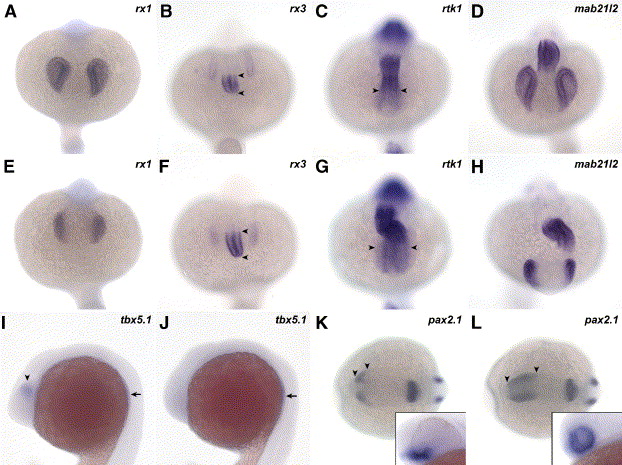
Fig. 5 The patterning of the eye is affected at the level of the RPE. Markers for the different axis of the eye were scored at 20 somites (A–J), 10 somites (K, L) or 30 hpf (insets in panels K, L) in wt (A–D, I, K) and chkw embryos (E–H, J, L). At the 20-somite stage, the expression domain of rx1 (A, E), rx3 (B, F) and mab21l2 (D, H) is reduced in the NR of chk. In contrast, the expression in the diencephalon of rtk1 is expanded into the NR compared to wt (arrowheads in panels G and C, respectively). Similarly, the expression pattern of rx3 is expanded in the hypothalamus (arrowheads in B and F). The ventral marker pax2.1 (K, L) is expanded into dorsal and posterior regions of the eye (arrowheads in K and L) at 10 somites. At 30 hpf, the expression domain of pax2.1 includes the entire NR in chk, while in wt it is restricted to the optic stalk region (insets in panels K, L). The expression of tbx5.1 is observed in the dorsal region of the eye (arrowhead in I), but it is lost in mutant embryos (J) at 20 somites. However, tbx5.1 is still present in the rudiments of the pectoral fins (arrows in I and J). The images in the figure are dorsal view, anterior downward (A–H); lateral view, anterior to the left (I, J); dorsal view, anterior to the left (K, L); and lateral view of a first plane of the eye (insets in panels K and L). Abbreviations like in Fig. 1.
Reprinted from Developmental Biology, 288(2), Rojas-Munoz, A., Dahm, R., and Nüsslein-Volhard, C., chokh/rx3 specifies the retinal pigment epithelium fate independently of eye morphogenesis, 348-362, Copyright (2005) with permission from Elsevier. Full text @ Dev. Biol.