
Fig. 10
- ID
- ZDB-IMAGE-070917-68
- Genes
- Publication
- Hamade et al., 2006 - Retinoic acid activates myogenesis in vivo through Fgf8 signalling
- All Figures
- Figures for Hamade et al., 2006
|
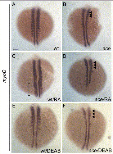
Fig. 10 Induction of myoD by RA requires a functional Fgf8. 10-somite stage embryos are displayed as in Fig. 7. (A–B) Untreated embryos. In ace, expression of myoD is reduced in the lateral aspect of somites, but remains expressed in a domain close to adaxial cells in anterior somites (arrowheads in panel B). (C–D) RA-treated embryos; expression of myoD is strongly induced in somites and psm of wild-type embryos (bracket in panel C). In ace, expression of myoD is weak in somites (arrowheads) and reduced in adaxial cells at the level of psm (bracket; D). (E–F) DEAB-treated embryos; expression of myoD is reduced in somites of wild-type embryos (E) compared to untreated wild-type (A). Reduction is stronger in somites of ace embryos (F; arrowheads) compared to DEAB-wild-type (E) and untreated-ace embryos (B). Scale bar: in panel A, 100 μm for panels A–F.
Reprinted from Developmental Biology, 289(1), Hamade, A., Deries, M., Begemann, G., Bally-Cuif, L., Genet, C., Sabatier, F., Bonnieu, A., and Cousin, X., Retinoic acid activates myogenesis in vivo through Fgf8 signalling, 127-140, Copyright (2006) with permission from Elsevier. Full text @ Dev. Biol.