
|
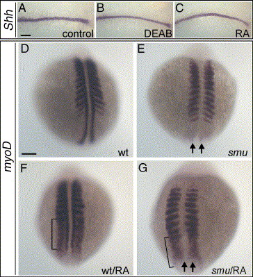
Fig. 7 Expression of myoD can be induced by RA in absence of Hh signalling. (A–C) 10-somite stage embryos displayed as in Fig. 2. At this stage, shh is expressed in embryo midline and no difference is observed upon DEAB or RA treatment. (D–G) Dorsal views of myoD expression in 10-somite stage embryos. (D, E) myoD fails to be expressed in adaxial cells in smu embryos (arrows in panel E). (F, G) In RA-treated embryos, myoD is induced in psm (bracket) in wild-types (F) and smu (G). Note that myoD remains absent from adaxial cells in RA-treated smu embryos (arrows). Scale bars: in panel A, 100 μm for panels A–C; in panel D, 100 μm for panels D–G.
Reprinted from Developmental Biology, 289(1), Hamade, A., Deries, M., Begemann, G., Bally-Cuif, L., Genet, C., Sabatier, F., Bonnieu, A., and Cousin, X., Retinoic acid activates myogenesis in vivo through Fgf8 signalling, 127-140, Copyright (2006) with permission from Elsevier. Full text @ Dev. Biol.