
|
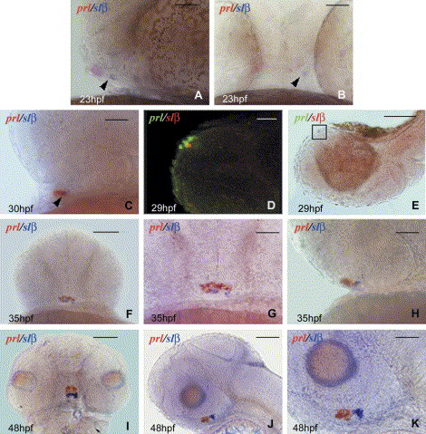
Fig. 1 Slβ expression pattern during embryogenesis. WT zebrafish embryos at 23 hpf (A,B), 29 hpf (D,E), 30 hpf (C), 35 hpf (F–H) and 48 hpf (I–K). (A,C,H,J,K) lateral view, anterior to the left; (B,F,G,I) frontal view, dorsal to the top; (D,E) lateral view, ventral to the top, anterior to the left; corresponding to the orientation in supplementary material S1. The scale bars in each picture correspond to 40 μm. Picture (B) corresponds to an assembly of two pictures: the same embryo focused in the medial part on prl and slβ signals, assembled with a picture of a more anterior plane with eyes in focus to be able to spot the morphology in comparison with the red and blue label. The arrows point at the slβ label. Prl (in red) is expressed anterior to slβ (in blue) at 23 hpf (A), in several cells (B), while slβ is detected only in one cell located on the left side of the embryo (B). At 30 hpf, slβ- and prl-expressing cells are intermingled (C). (D) Prl and slβ are expressed in different cells. Merger of red and green fluorescence in confocal microscopy: A 29 hpf embryo was labeled by combined fluorescent in situ hybridization: red fluorescence for slβ and green for prl. (E) classical microscopy picture showing the orientation of the embryo represented in (D) and in supplementary material S1, lateral view, ventral to the top, anterior to the left. Note the red slβ label. The square outlines the enlarged portion in (D) and S1. (F–K) The blue label corresponds to slβ, the red label to prl. Slβ expression increases at 35 hpf (F–H) through 48 hpf (I–K) and the expressing cells move posterior relative to the prl domain.
Reprinted from Gene expression patterns : GEP, 6(2), Lopez, M., Nica, G., Motte, P., Martial, J.A., Hammerschmidt, M., and Muller, M., Expression of the somatolactin beta gene during zebrafish embryonic development, 156-161, Copyright (2006) with permission from Elsevier. Full text @ Gene Expr. Patterns