
|
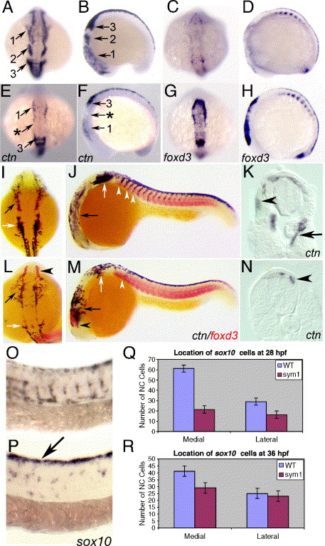
Fig. 7 Abnormal cell migration in sym1 mutant embryos. Wild-type (A–D) and sym1 mutant (E–H) embryos at the 12-somite stage, showing dorsal (A, E, C, G) and lateral (B, F, D, H) views. ctn expression reveals migration of the three cranial neural crest streams in wild-type embryos (A–B, numbered arrows). In sym1 embryos, ctn expression in cranial neural crest is severely reduced in the 1st neural crest stream (E, F) and almost absent in the 2nd stream (arrow with asterisk in E, F). ctn-positive cells of the 3rd stream are present in sym1 mutants, but only a few cells migrate ventrally (compare F with B). At the same stage, foxd3 expression is normally rapidly downregulated in wild-type embryos upon migration (C–D), but persists in sym1 mutants (G–H), showing that the cranial premigratory crest is present, but does not migrate. Wild-type (I–J) and sym1 mutant (L–M) embryos double labeled with ctn (black) and foxd3 (red) at the 26-somite stage, showing dorsal (I, L) and lateral (J, M) views. At this stage, ctn is expressed throughout the head in deep ventral positions of wild-type embryos (I, J, black arrow); however, in sym1 mutants, ctn-positive cells fail to migrate ventrally, and spread out more laterally (L, M, black arrow). The expression of ctn is also strongly reduced in the region of the hindbrain posterior to the otic vesicle in sym1 mutants (L–M, white arrows), while foxd3 expression remains high in the anterior-most neural crest cells (L, M, black arrowhead). Numerous, ctn-expressing trunk neural crest cells are seen migrating in wild-type embryos at the 26-somite stage (J; white arrowhead), but few ctn-positive cells are seen in sym1 embryos (M; white arrowhead). Transverse sections at the anterior trunk level of 24 hpf wild-type (K) and sym1 mutant (N) embryos, showing ctn expression in both dorsal-laterally (K, arrowhead) and medial (K, arrow) migrating neural crest cells in wild-type embryos. In sym1 mutants, migration of ctn-expressing cells are delayed, and found only in dorsal positions at this stage (N, arrowheads). Lateral views of sox10-expressing neural crest cells in wild-type (O) and sym1 (P) embryos at 24 hpf. In wild-type embryos, robust sox10 expression is evident in migrating neural crest cells; however, in sym1 mutants most of the sox10-expressing cells are still on the dorsal neural tube (P, arrow). Quantitation of sox10-positive cells located along each migration pathway at 28 hpf (Q) and 36 hpf (R) in wild-type (blue) and sym1 mutants (red), shows that migration along the medial pathway is more severely affected in sym1 mutants.
Reprinted from Developmental Biology, 292(1), Stewart, R.A., Arduini, B.L., Berghmans, S., George, R.E., Kanki, J.P., Henion, P.D., and Look, A.T., Zebrafish foxd3 is selectively required for neural crest specification, migration and survival, 174-188, Copyright (2006) with permission from Elsevier. Full text @ Dev. Biol.