
|
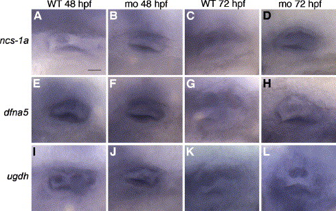
Fig. 4 Expression of semicircular canal markers in α1a.2 morphant ears. All panels are lateral views with anterior to the left. Embryos were injected with 2 ng of α1a.2 MO-1 at the one-cell stage. (A–D) ncs-1a staining in the otic vesicle (OV) of (A) 48 hpf wild type (WT) embryo, (B) 48 hpf morphant, (C) 72 hpf WT embryo, and (D) 72 hpf morphant. (E–H) dfna5 staining in OV of (E) 48 hpf WT embryo, (F) 48 hpf morphant, (G) 72 hpf WT embryo, and (H) 72 hpf morphant. (I–L) ugdh staining in OV of (I) 48 hpf WT embryo, (J) 48 hpf morphant, (K) 72 hpf WT embryo, and (L) 72 hpf morphant. mo, morphant. Scale bar: 25 μm.
Reprinted from Developmental Biology, 294(1), Blasiole, B., Canfield, V.A., Vollrath, M.A., Huss, D., Mohideen, M.A., Dickman, J.D., Cheng, K.C., Fekete, D.M., and Levenson, R., Separate Na,K-ATPase genes are required for otolith formation and semicircular canal development in zebrafish, 148-160, Copyright (2006) with permission from Elsevier. Full text @ Dev. Biol.