
|
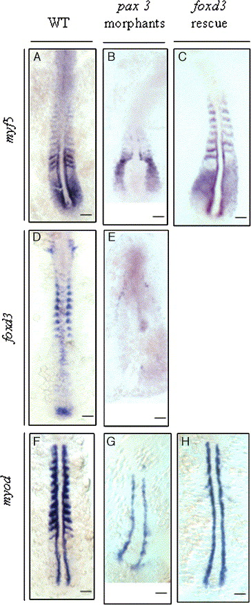
Fig. 8 The effect of inhibiting Pax3 protein synthesis on the expression of foxd3, myf5, and myod in the zebrafish embryos at 5 to 8 somites. Probes were used to detect myf5 (A, B, C), foxd3 (D, E), and myod (F, G, H) in the wild-type embryos (A, D, F), the pax3-MO1-injected (6 ng) embryos (B, E, G), and the embryos co-injected with 6 ng of pax3-MO1 and 25 pg of foxd3 mRNA (C, H). (B) In pax3-MO1-injected embryos, myf5 expression was restricted in the presomitic mesoderm (PSM) and weak in somites 0 and -1 and in the adaxial cells on the sides of somites 0 and -1. (E) The shape of the neural plate became abnormal in the pax3-MO1 morphants, and foxd3 expression was weak in their neural fold and tail bud. (G) Meanwhile, myod expression was down-regulated in the somites, but it was expressed in adaxial cells. In the rescue experiment, co-injection of foxd3 mRNA and pax3-MO1 restored myf5 expression in the somites (C), whereas the myod expression was not rescued (H). Scale bars: 100 μm.
Reprinted from Developmental Biology, 290(2), Lee, H.C., Huang, H.Y., Lin, C.Y., Chen, Y.H., and Tsai, H.J., Foxd3 mediates zebrafish myf5 expression during early somitogenesis, 359-372, Copyright (2006) with permission from Elsevier. Full text @ Dev. Biol.