
|
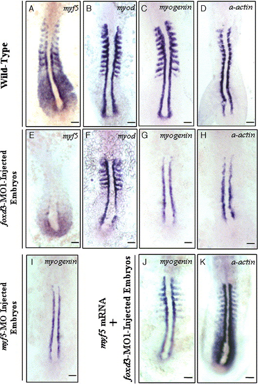
Fig. 7 Effect of inhibiting Foxd3 protein synthesis on myf5, myod, myogenin, and α-actin expression in embryos with 10–12 somites. In foxd3-MO1-injected (10 ng) embryos, myf5 expression in the somites, adaxial cells, and presomitic mesoderm was reduced dramatically (A vs. E) but myod expression was unchanged (B vs. F). In foxd3-MO1-injected embryos, the expression of myogenin (C vs. G) and α-actin (D vs. H) was abolished in the somites, except in adaxial cells. Embryos injected with 4 ng of myf5-MO exhibited reduced myogenin expression in the somites, a finding similar to the defective phenotype of foxd3 morphants (I vs. G). Co-injection of 600 ng/μl myf5-capped mRNA rescued expression of myogenin (J) and α-actin (K) in foxd3-MO1-injected embryos. Scale bars: 100 μm.
Reprinted from Developmental Biology, 290(2), Lee, H.C., Huang, H.Y., Lin, C.Y., Chen, Y.H., and Tsai, H.J., Foxd3 mediates zebrafish myf5 expression during early somitogenesis, 359-372, Copyright (2006) with permission from Elsevier. Full text @ Dev. Biol.