
|
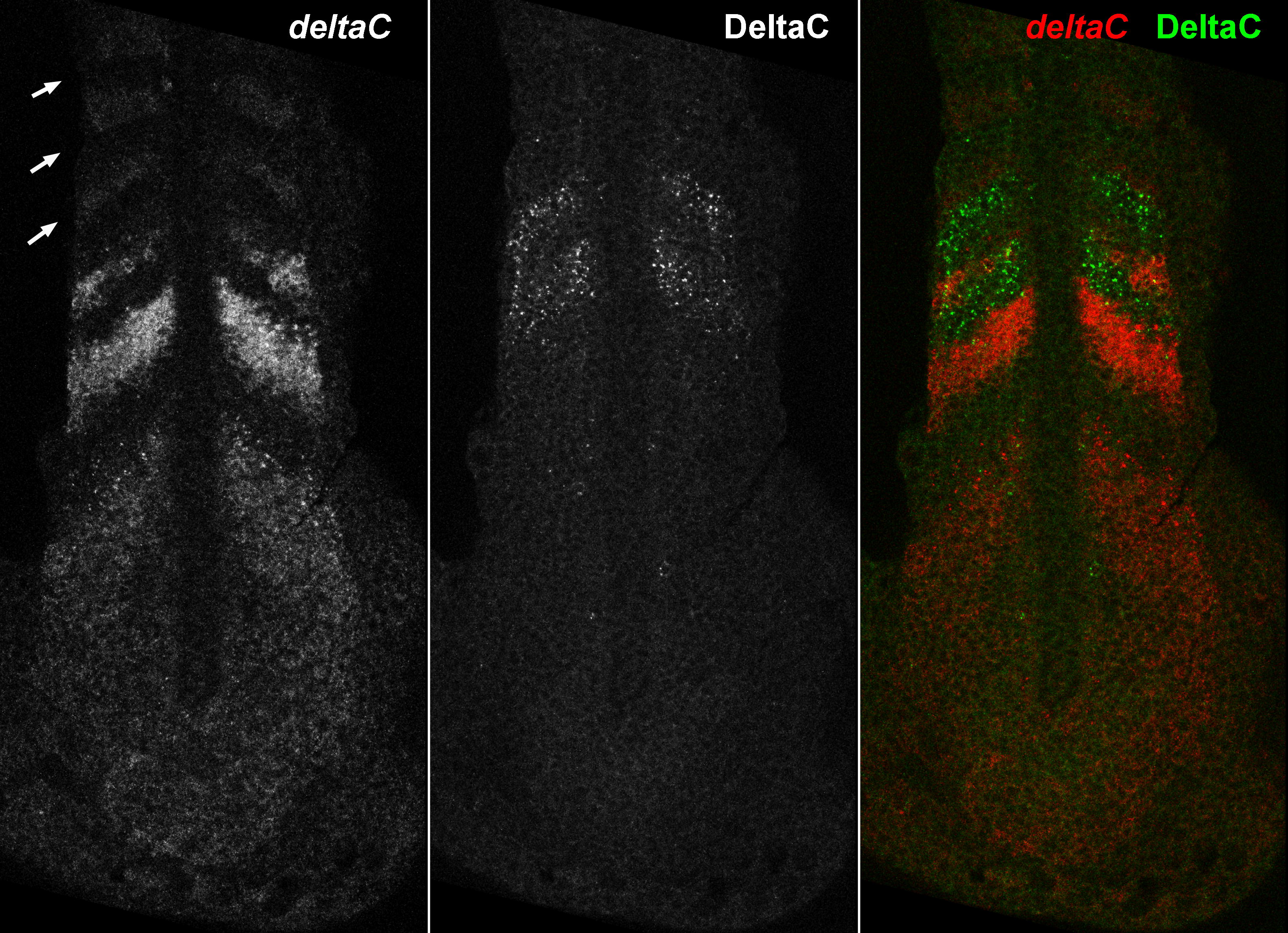
Fig. 5 The Pattern of deltaC Transcripts Compared with the Pattern of DeltaC Protein in a Doubly Stained Specimen. The left-hand panel shows the distribution of deltaC mRNA, as revealed by ISH; arrows indicate the three most recently formed somites. The middle panel shows the distribution of DeltaC protein in the same optical section, immunostained with the zdc2 monoclonal antibody. The right-hand panel shows the two patterns superimposed in the doubly stained specimen. The protein pattern is shifted by almost exactly one somite width relative to the mRNA pattern; one somite cycle time earlier, the cells currently displaying peak levels of protein would have been in the locations of the cells currently expressing the peak levels of mRNA and would have been expressing those levels of mRNA themselves. Thus there is a delay of approximately one somite cycle time (30 min) from the accumulation of the mRNA in the cell to the accumulation of the protein translated from it.