
|
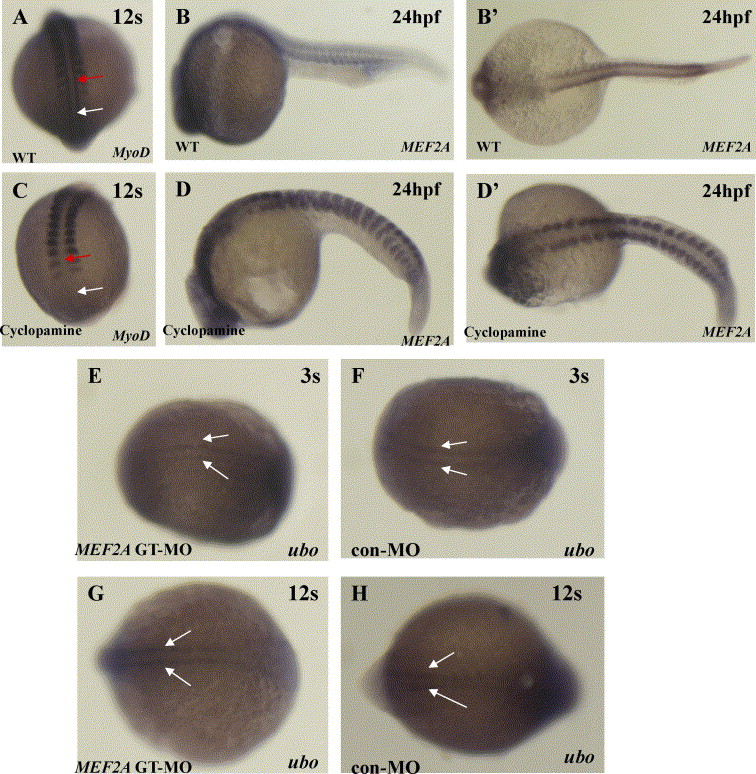
Fig. 5 Hedgehog signaling negatively regulates MEF2A expression. (A and C) Dorsal views of whole-mount embryos at the 12-somite stage to show MyoD expression. Wild-type embryos (A, n = 15 embryos) exhibit adaxial MyoD expression throughout the somitic (red arrow) and presomitic (white arrow) mesoderm, while embryos (C, n = 10 embryos) treated with 25 μM cyclopamine at 6 hpf lack expression in the somitic (red arrow) and in the presomitic (white arrow) mesoderm. (B and D) (B, n = 15 embryos; D, n = 10 embryos) Lateral views of whole-mount embryos with the head to the left at 24 hpf. (B′ and D′) (B, n = 15 embryos; D, n = 10 embryos) Dorsal views of whole-mount embryos with the head to the left at 24 hpf. MEF2A expression is increased in the somites of cyclopamine-treated embryos. (E–H) MEF2A is not required for ubo transcription. Dorsal views of whole-mount embryos at the 3-somite stage (E and F) (E, n = 20 embryos; F, n = 20 embryos) and the 12-somite stage (G and H) (G, n = 20 embryos; H, n = 20 embryos) to show ubo expression. (F) At the 3-somite stage, ubo is expressed in the adaxial cells (white arrow). (H) At the 12-somite stage, ubo is expressed prominently in the newly formed posterior somites (white arrow). Expression in MEF2A morphants of ubo is similar to that observed in control embryos (E and G).
Reprinted from Mechanisms of Development, 123(10), Wang, Y., Qian, L., Dong, Y., Jiang, Q., Gui, Y., Zhong, T.P., and Song, H., Myocyte-specific enhancer factor 2A is essential for zebrafish posterior somite development, 783-791, Copyright (2006) with permission from Elsevier. Full text @ Mech. Dev.