
Image
|
Figure Caption
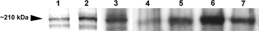
Fig. 4 Robo3 protein expression. In vitro translated, Myc-epitope tagged Robo3var1 and Robo3var2 proteins were identified using an anti-Myc antibody (Lanes 1 and 2, respectively). Robo3var2-Myc (Lane 3) and Robo3var1-Myc (not shown) were also recognized by anti-Robo3 antiserum. Robo3 proteins from zebrafish embryo extracts, corresponding to the same size as that of in vitro translated proteins, were also identified by anti-Robo3 antiserum (Lanes 4–7). Robo3 proteins were observed as early as 100% epiboly (Lane 4) and continued to be expressed at 11, 12, and 16–18 hpf (Lanes 5–7, respectively).
Figure Data
Acknowledgments
This image is the copyrighted work of the attributed author or publisher, and
ZFIN has permission only to display this image to its users.
Additional permissions should be obtained from the applicable author or publisher of the image.
Reprinted from Mechanisms of Development, 122(10), Challa, A.K., McWhorter, M.L., Wang, C., Seeger, M.A., and Beattie, C.E., Robo3 isoforms have distinct roles during zebrafish development, 1073-1086, Copyright (2005) with permission from Elsevier. Full text @ Mech. Dev.