
|
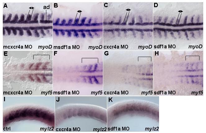
Fig. 2 Cxcr4 signaling is required for transcription of myogenic genes in the paraxial mesoderm. Dorsal (A-H) and lateral views (I-K). 13 h embryos hybridized with (A-D) myoD, (E-H) myf5 and (I-K) mylz2 riboprobes. (A,B) mcxcr4a (n = 52/52) and msdf1a morphants (n = 50/61) as controls. Embryos show expression pattern of myoD. (C) cxcr4a (n = 48/50) morphants show myoD transcription is reduced in the paraxial mesoderm, while expression in adaxial cells is unchanged. (D) sdf1a (n = 50/61) morphants show similar reduction of myoD in the paraxial cells but not adaxial cells. Black lines and arrows indicate size of expression domain. In addition, intensity of staining in lateral mesoderm is substantially reduced. (E,F) mcxcr4a (n = 36/36) and msdf1a (n = 49/58) morphants as controls. Embryos show characteristic expression pattern of myf5 in the adaxial cells, somitic mesoderm and presomitic mesoderm. (G) cxcr4a (n = 43/47) morphants have myf5 reduced in both somites and forming somites. (H) sdf1a (n = 56/71) morphants cause similar effects to Cxcr4a knock down. Black brackets indicate a region where pattern and intensity of myf5 staining in the newly formed and forming somites were reduced. (I-K) Control (n = 30). Reduced mylz2 transcription in cxcr4a (n = 30) and sdf1a (n = 30) morphants. Abbreviation: ad – adaxial cells.