
|
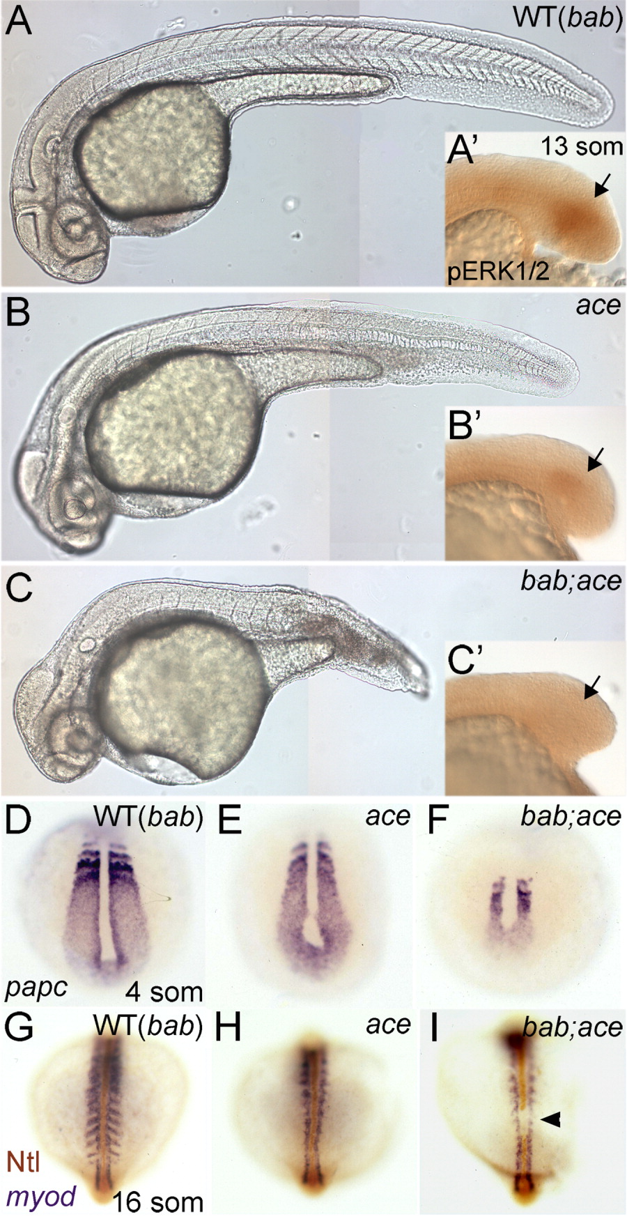
Fig. 5 rerea interacts genetically with fgf8 in posterior mesoderm development. A-C: Lateral views of live 24 hpf embryos with genotypes in the upper right corner. A′-C′: Inset panels show the corresponding lateral views of diphosphorylated ERK1&2 (pERK) immunostained tail buds at 13 somites. Arrows denote the position of pERK staining. D-I: Whole-mount in situ hybridizations of papc at 4 somites (D-F) or myod with Ntl immunostains at 16 somites (G-I) shown in dorsal view with genotypes shown in the upper right corner. The arrowhead in I marks a gap in Ntl staining. "WT(bab)" notes that there is no observable difference between wild-type (WT) and bab embryos. ace shows a slight reduction in the amount of posterior mesoderm (B) and presomitic mesoderm as seen by papc expression at four somites (E), while bab;ace (C,F) shows a severe reduction in comparison to wild-type or bab (A,D).