
|
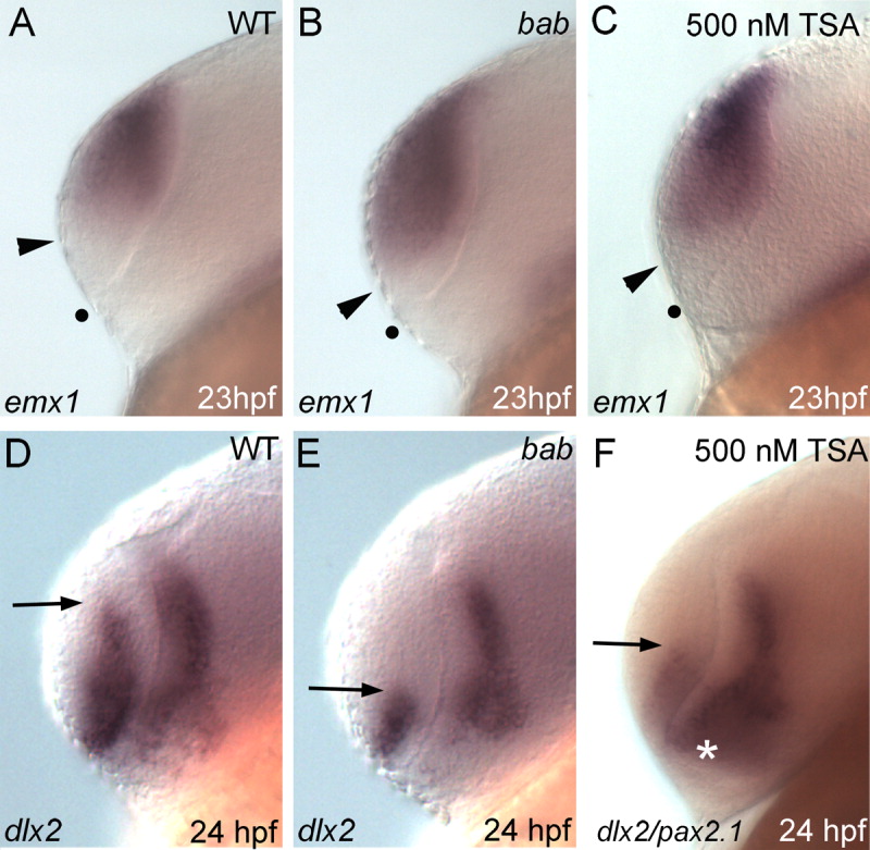
Fig. 3 Disruption of REREa or histone deacetylase (HDAC) activity causes a dorsalization of the telencephalon. A-F: All panels show lateral views of the telencephalon in whole-mount in situ hybridizations on wild-type (WT, A,D), bab (B,E) or Trichostatin A (TSA) -treated (C,F) embryos. Probes are denoted in the lower left corner with stages in the lower right. A-C: emx1 expression extends ventrally from the dorsal telencephalon to the arrowhead and is expanded in bab and TSA-treated embryos. The dot marks the ventral telencephalic boundary. D-F: This results in a reduction of dlx2 expression in the telencephalon. Arrows mark the dorsal limit of dlx2 expression. pax2.1 expression (white asterisk) in F overlaps the diencephalic expression of dlx2, but is not expressed in the telencephalon.