
Image
|
Figure Caption
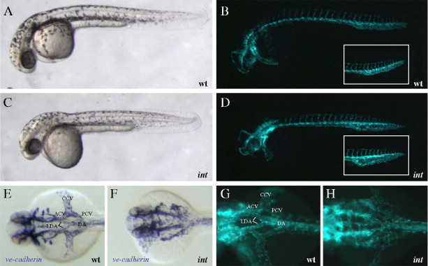
Fig. 6 intersection regulates vascular patterning. (A–D) Lateral bright-field and epifluorescent micrographs of 32 hpf embryos. Vascular patterning defects can be observed by visualizing Tg(flk1:EGFP)s843 expression, but not via bright-field microscopy. ints413 mutants fail to connect the lateral dorsal aortae (LDA) to form the dorsal aorta (DA), and the anterior and posterior cardinal veins (ACV and PCV) to the common cardinal vein (CCV). (E–H) Dorsal views of ve-cadherin and Tg(flk1:EGFP)s843 expression. ints413 mutant embryos exhibit a pronounced dilation of the PCV (inset in panel D).
Figure Data
Acknowledgments
This image is the copyrighted work of the attributed author or publisher, and
ZFIN has permission only to display this image to its users.
Additional permissions should be obtained from the applicable author or publisher of the image.
Reprinted from Developmental Biology, 307(1), Jin, S.W., Herzog, W., Santoro, M.M., Mitchell, T.S., Frantsve, J., Jungblut, B., Beis, D., Scott, I.C., D'Amico, L.A., Ober, E.A., Verkade, H., Field, H.A., Chi, N.C., Wehman, A.M., Baier, H., and Stainier, D.Y., A transgene-assisted genetic screen identifies essential regulators of vascular development in vertebrate embryos, 29-42, Copyright (2007) with permission from Elsevier. Full text @ Dev. Biol.