
|
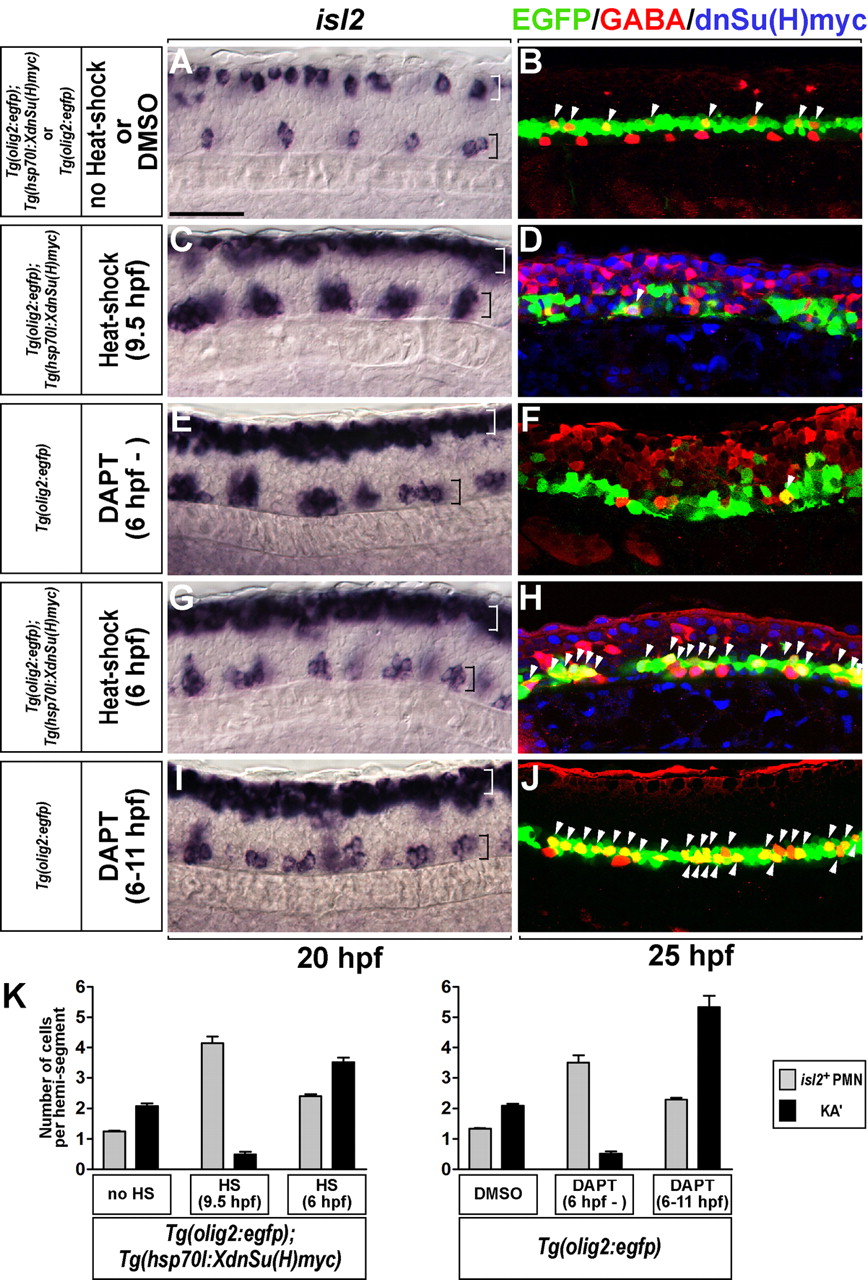
Fig. 4 Transient inactivation of Notch signaling produces excess PMNs and KA' interneurons. (A-J) Lateral views of the spinal cord of zebrafish embryos at the 6- to 12-somite region, with anterior to the left and dorsal at the top. All embryos carried the Tg(olig2:egfp) transgene. (C,D,G,H) Tg(olig2:egfp);Tg(hsp70l:XdnSu(H)myc) double-transgenic embryos. (A,C,E,G,I) isl2 expression at 20 hpf. White and black brackets mark dorsal and ventral spinal cord, respectively. (B,D,F,H,J) KA' cells (arrowheads) were detected by anti-GABA antibody (red) within EGFP+ cells (green) at 25 hpf. (A,B) Control embryos. (C,D) Induction of XdnSu(H)Myc by heat shock at 9.5 hpf resulted in excess PMNs with reduction of KA' interneurons (arrowhead). XdnSu(H)Myc protein was detected by anti-Myc antibody (blue). (E,F) Embryos incubated continuously with DAPT had excess PMNs and a deficit of KA' interneurons (arrowhead). (G,H) Double-transgenic embryos heat shocked at 6 hpf formed excess PMNs (black bracket) and KA' interneurons (arrowheads). (I,J) Embryos incubated with DAPT from 6 to 11 hpf had excess PMNs (black bracket) and KA' cells (arrowheads). (K) Mean of the number of PMNs (gray bar) and KA' interneurons (black bar) per hemisegment (n=7) in A-J. Error bars represent s.e.m. Scale bar: 50 µm.