
|
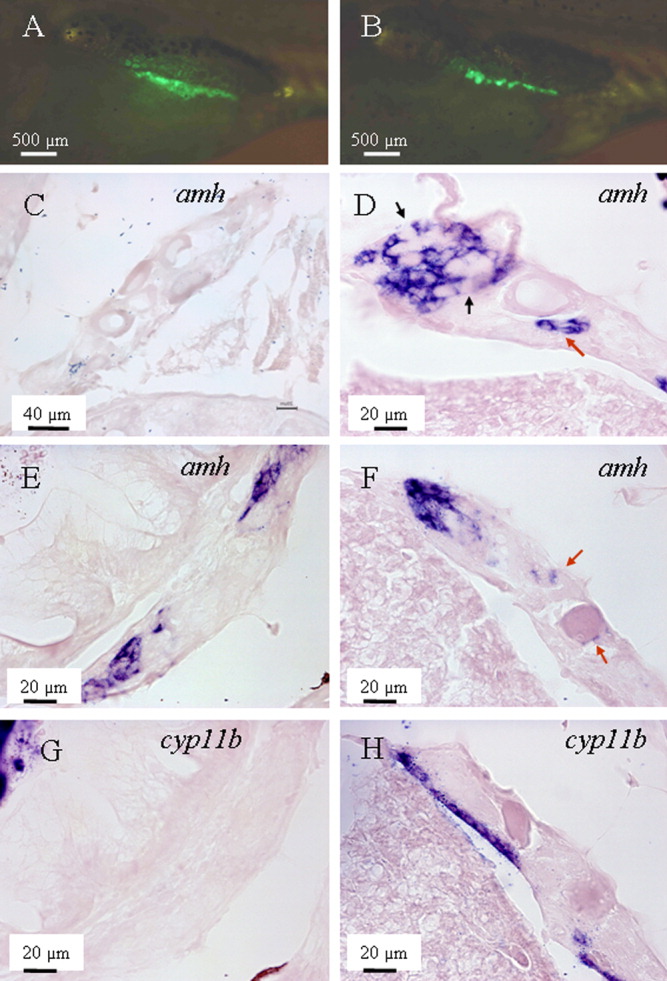
Fig. 6 amh was expressed earlier than cyp11b during gonadal transformation. A, C, E, G: A male zebrafish at the early stage of transformation (35 days postfertilization [dpf]). B, D, F, H: Another male of the same age at advanced stage of gonad transformation. A,B: In vivo recording of enhanced green-fluorescent protein (EGFP) level in the gonad. C,E: In the early transforming gonads, amh could not be detected in the gonadal regions full of oocytes (C), but became detectable when the number of oocytes decreased (E). G: At the same time, cyp11b could not be detected anywhere during that time period. D,F: In the gonads at advanced stages of transformation, amh was expressed in most regions analyzed, localized to the somatic cells surrounding new germ cells (likely spermatogonia; black arrows on D) or close to the degenerating oocytes (red arrows on D and F). H:cyp11b only started to be expressed in some regions of the same gonad. It was expressed first on the edge of the gonads, then later also within the gonads (not shown). Notes: (1) due to variability of the transformation process male individuals of the same age might be at different stages of their gonad development; (2) sections shown on E and G were adjacent, and so were those on F and H.