
|
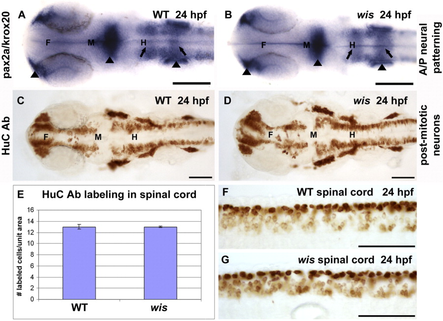
Fig. 6 Neuronal determination is normal in whitesnake mutants. A,B: In situ hybridization for pax2a (labeling nasal placodes, midbrain-hindbrain boundary, and otic vesicles, arrowheads) and krox20 (labeling rhombomeres 3 and 5, arrows) show similar staining patterns in wild-type and mutant. C,D: Immunohistochemistry for HuC, a marker for postmitotic neurons, shows identical patterns between wild-type and mutant. E-G: This finding has been quantified in graph E, which depicts the number of HuC-labeled cells per unit area in the spinal cords of wild-type and wis (F,G). F, forebrain; M, midbrain; H, hindbrain. Scale bar = 100 μm.