
|
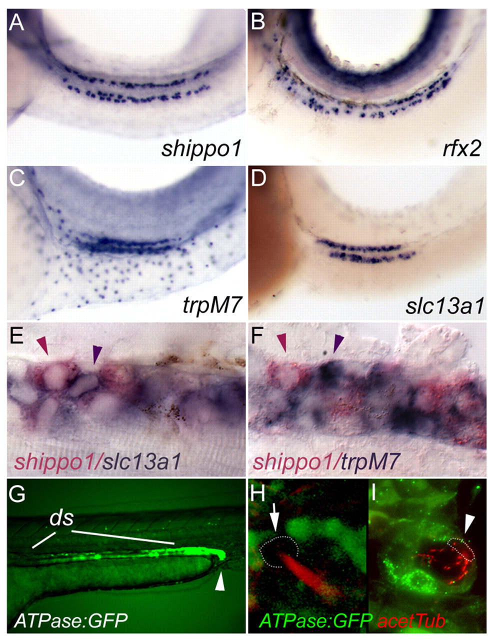
Fig. 2 MCCs and transporting epithelia co-exist as separate cell types in the early distal segment of the pronephric nephron. (A) Expression of the axonemal-sheath gene shippo1 in the pronephros of a 34 hpf embryo. (B) Expression of the transcription factor rfx2 in individual pronephric cells. (C) Expression of the cation transporter trpM7 in the early distal segment of the pronephric nephron. (D) Expression of the sodium-sulfate co-transporter slc13a1 in the early distal segment. (E) Double in situ hybridization of the MCC marker shippo1 (red) and slc13a1 (purple) in distinct but adjacent cells of the early distal segment in a 34 hpf embryo. (F) Double in situ hybridization of shippo1 and trpM7 in early distal segment cells. (G) Expression of GFP from the Na,K-ATPase alpha a1A4 subunit promoter is uniform in the most caudal nephron segment (arrowhead) and heterogeneous in the distal nephron. (H,I) Double staining for GFP and acetylated tubulin (red) in confocal z-series projections (H) and in histological sections (I) shows that the Na,K-ATPase alpha a1A4 promoter is not active in MCCs (arrows, arrowhead; cell bodies are outlined with dashed line).