
Fig. 3
|
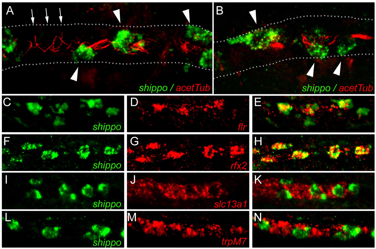
Fig. 3 Confocal fluorescent in situ hybridization analysis of ion transporter and ciliogenic gene expression in the pronephros. (A,B) Double staining for shippo1 mRNA (in situ hybridization; green) and acetylated tubulin (immunofluorescence; red) in confocal projections reveals several examples of cilia bundles emanating from shippo1-positive cells (arrowheads) into the lumen; single cilia emanate from shippo1-negative cells (small arrows). Dotted lines outline the pronephros; posterior is to the right. (C-E) Co-expression of shippo1 (C; green) and fleer (D; red) in single cells of the pronephric tubules. (E) Merged image. (F-H) Co-expression of shippo1 (F; green) and rfx2 (G; red) in single cells of the pronephric tubules. (H) Merged image. (I-K) Expression of shippo1 (I; green) and slc13a1 (J; red) in distinct but adjacent cells of the pronephric tubules. (K) Merged image. (L-N) Expression of shippo1 (L; green) and trpM7 (M; red) in distinct but adjacent cells of the pronephros. (N) Merged image.