
|
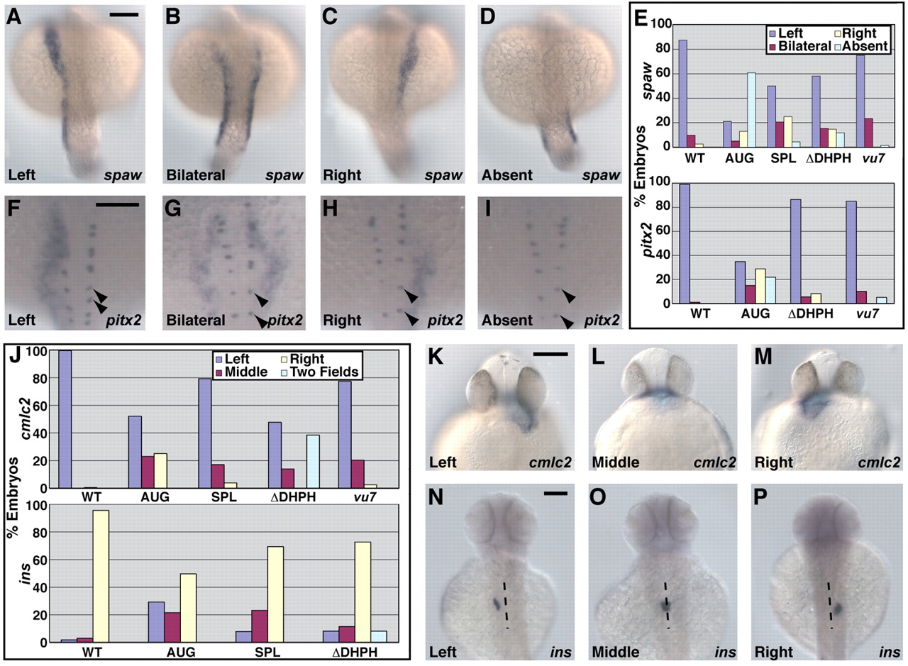
Fig. 3 Loss of Arhgef11 function leads to abnormal expression of laterality markers. (A-D) Expression of spaw in 19- to 22-somite stage (∼19 hpf) embryos detected by in situ hybridization with antisense RNA probe. Scale bar, 150 μm. (E) Bar graph of the percentage of WT and treated embryos exhibiting the expression patterns for spaw shown in A-D, or pitx2 shown in F-I. (F-I) Expression of pitx2 in 22- to 25-somite stage (∼21 hpf) embryos detected by in situ hybridization with antisense RNA probe. Expression of pitx2 in Rohon Beard cells (marked by arrowheads) is not disrupted. Scale bar, 150 μm. (J) Graphical representation of the percentage of embryos exhibiting the expression patterns for cmlc2 shown in K-M or for ins shown in N-P. (K-M) Expression of cmlc2 in ∼33 hpf embryos detected by in situ hybridization with antisense RNA probe. Scale bar, 150 μm. (N-P) Expression of ins in ∼53 hpf embryos detected by in situ hybridization with antisense RNA probe. A dotted line marks the approximate midline. Scale bar: 150 μm.