
|
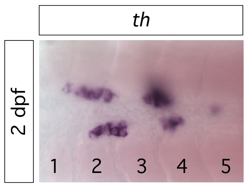
Fig. S1 Morphology of cervical sympathetic ganglia in 2 dpf zebrafish embryos. After whole-mount in situ hybridisation for th as a marker for sympathetic neurons, dorsal hindbrain/spinal cord and yolk sac were dissected and viewed from dorsal using 40× objective. Please note individual cells with nuclei spared from the in situ staining reaction in the plane of focus and other cells that give a blurred appearance, as they are out of focus. The embryo shown was selected for the number of cells within one focus plane. Somite numbers are indicated to document that cervical sympathetic ganglion cells are mainly located betwen somites 1 and 4.