
|
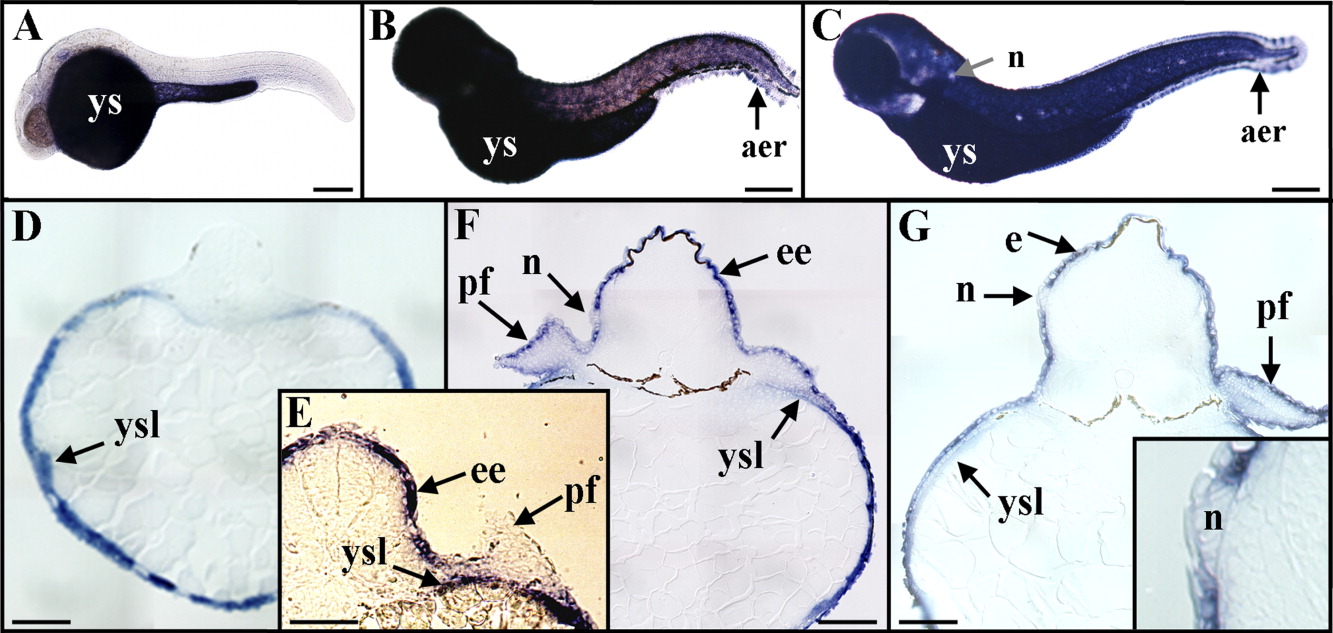
Fig. 2 Expression of rbp4 during zebrafish embryonic and early larval development. A-C: Whole-mount in situ hybridizations, by using digoxigenin-labeled antisense riboprobes, are shown in lateral views and anterior to the left at 24 hours postfertilization (hpf; A), 48 hpf (B), and 72 hpf (C). D-G: Transverse sections after in situ hybridizations at 24 hpf (D), 42 hpf (E), 48 hpf (F), and 72 hpf (G). A: By 24 hpf, rbp4 hybridization signal was found in the yolk sac. D: Histological sections demonstrated that rbp4 transcripts were restricted to the yolk syncytial layer. E: By 42 hpf, an epidermal expression of rbp4 began, but no transcripts were detected in pectoral fin-bud epidermis. B,C,F,G: By 48 hpf (B,F) and 72 hpf (C,G, and insert), rbp4 transcripts were detected in the embryonic epidermis, in pectoral fin-bud epidermis, in the yolk syncytial layer, in dorsal and ventral apical ectodermal ridges, but not in neuromasts. aer, apical ectodermal ridge; e, epidermis; ee, embryonic epidermis; n, neuromast; tb, tail bud; pf, pectoral fin buds; ys, yolk sac; ysl, yolk syncytial layer. Scale bars = 200 μm.