
|
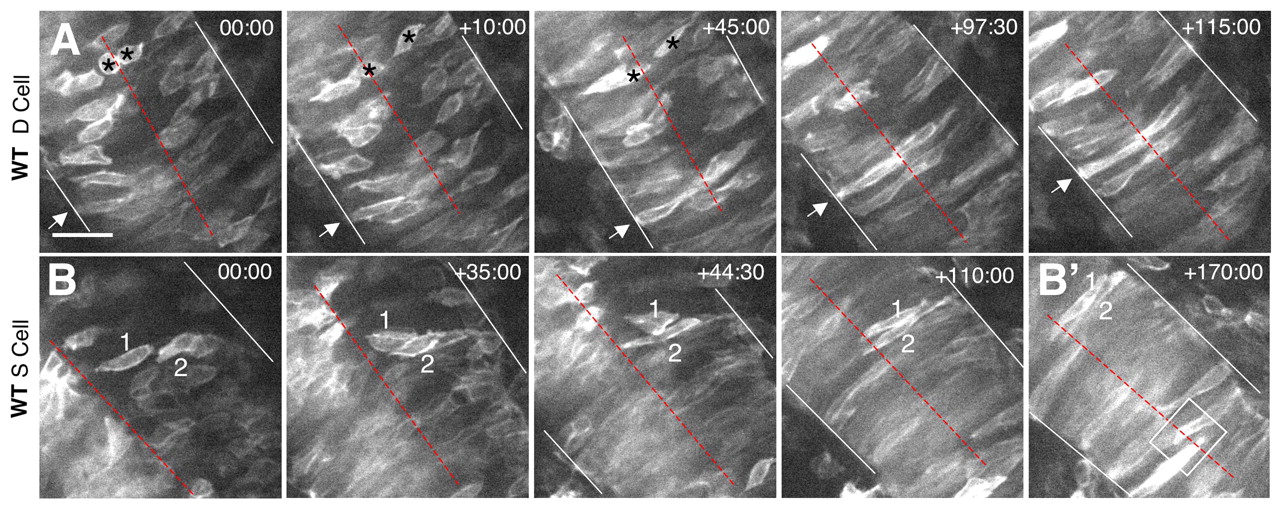
Fig. 3 Time-lapse imaging of wild-type cells. (A,B) Selected frames from time-lapse confocal movies are shown for a deep (A) and two superficial (B) cells. Time elapsed (in minutes) from the first frame is indicated in the upper right corner. mGFP-labeled embryos were imaged in the hindbrain region, from a dorsal view, beginning at approximately 2-3 som and extending through 6-7 som. Dotted red lines indicate the midline of the neural keel/rod, white lines mark the edges of the neural keel/tube, arrows and numbers indicate individual cells identified in multiple frames, asterisks in A indicate two daughter cells derived from a recent cell division, and the box in B' shows interdigitation of cells at the midline. Scale bar: 100 μm.